 Movie
Movie Controller


+ Open data

- Basic information
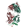
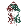
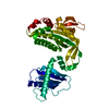
| Entry | Database: PDB / ID: 6rcq | ||||||
|---|---|---|---|---|---|---|---|
| Title | PfRH5-binding monoclonal antibody R5.011 | ||||||
 Components Components |
| ||||||
 Keywords Keywords | IMMUNE SYSTEM / Plasmodium falciparum Erythrocyte invasion Potentiating antibody Human monoclonal antibody | ||||||
| Function / homology | Immunoglobulins / Immunoglobulin-like / Sandwich / Mainly Beta / ACETATE ION / CACODYLATE ION Function and homology information Function and homology information | ||||||
| Biological species |  Homo sapiens (human) Homo sapiens (human) | ||||||
| Method |  X-RAY DIFFRACTION / SYNCHROTRON / MOLECULAR REPLACEMENT / Resolution: 2.28 Å X-RAY DIFFRACTION / SYNCHROTRON / MOLECULAR REPLACEMENT / Resolution: 2.28 Å | ||||||
 Authors Authors | Alanine, D.W.G. / Draper, S.J. / Higgins, M.K. | ||||||
| Funding support |  United Kingdom, 1items United Kingdom, 1items
| ||||||
 Citation Citation | Journal: Cell / Year: 2019 Title: Human Antibodies that Slow Erythrocyte Invasion Potentiate Malaria-Neutralizing Antibodies. Authors: Alanine, D.G.W. / Quinkert, D. / Kumarasingha, R. / Mehmood, S. / Donnellan, F.R. / Minkah, N.K. / Dadonaite, B. / Diouf, A. / Galaway, F. / Silk, S.E. / Jamwal, A. / Marshall, J.M. / Miura, ...Authors: Alanine, D.G.W. / Quinkert, D. / Kumarasingha, R. / Mehmood, S. / Donnellan, F.R. / Minkah, N.K. / Dadonaite, B. / Diouf, A. / Galaway, F. / Silk, S.E. / Jamwal, A. / Marshall, J.M. / Miura, K. / Foquet, L. / Elias, S.C. / Labbe, G.M. / Douglas, A.D. / Jin, J. / Payne, R.O. / Illingworth, J.J. / Pattinson, D.J. / Pulido, D. / Williams, B.G. / de Jongh, W.A. / Wright, G.J. / Kappe, S.H.I. / Robinson, C.V. / Long, C.A. / Crabb, B.S. / Gilson, P.R. / Higgins, M.K. / Draper, S.J. | ||||||
| History |
|
- Structure visualization
| Structure viewer | Molecule: MolmilJmol/JSmol |
|---|
- Downloads & links
-Download
| PDBx/mmCIF format | 6rcq.cif.gz | 181.8 KB | Display | PDBx/mmCIF format |
|---|---|---|---|---|
| PDB format | pdb6rcq.ent.gz | 143.5 KB | Display | PDB format |
| PDBx/mmJSON format | 6rcq.json.gz | Tree view | PDBx/mmJSON format | |
| Others |  Other downloads Other downloads |
-Validation report
| Arichive directory | https://data.pdbj.org/pub/pdb/validation_reports/rc/6rcqftp://data.pdbj.org/pub/pdb/validation_reports/rc/6rcq | HTTPS FTP |
|---|
-Related structure data


-Links
 PDBj
PDBj


- Assembly
| Deposited unit | 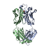
| ||||||||
|---|---|---|---|---|---|---|---|---|---|
| 1 |
| ||||||||
| Unit cell |
|
-Components
| #1: Antibody | Mass: 25685.484 Da / Num. of mol.: 1 Source method: isolated from a genetically manipulated source Source: (gene. exp.) Homo sapiens (human) / Production host: Homo sapiens (human) |
|---|---|
| #2: Antibody | Mass: 23065.367 Da / Num. of mol.: 1 Source method: isolated from a genetically manipulated source Source: (gene. exp.) Homo sapiens (human) / Production host: Homo sapiens (human) |
| #3: Chemical | ChemComp-ACT /   Mass: 59.044 Da / Num. of mol.: 1 / Source method: obtained synthetically / Formula: C2H3O2 Mass: 59.044 Da / Num. of mol.: 1 / Source method: obtained synthetically / Formula: C2H3O2 |
| #4: Chemical | ChemComp-CAC /   Mass: 136.989 Da / Num. of mol.: 1 / Source method: obtained synthetically / Formula: C2H6AsO2 Mass: 136.989 Da / Num. of mol.: 1 / Source method: obtained synthetically / Formula: C2H6AsO2 |
| #5: Water | ChemComp-HOH /  Mass: 18.015 Da / Num. of mol.: 238 / Source method: isolated from a natural source / Formula: H2O Mass: 18.015 Da / Num. of mol.: 238 / Source method: isolated from a natural source / Formula: H2O |
| Has protein modification | Y |
-Experimental details
-Experiment
| Experiment | Method: X-RAY DIFFRACTION / Number of used crystals: 1 |
|---|
- Sample preparation
| Crystal | Density Matthews: 2.7 Å3/Da / Density % sol: 54.46 % |
|---|---|
| Crystal grow | Temperature: 298 K / Method: vapor diffusion, sitting drop Details: 0.16 M ZnAc, 0.108 M Na cacodylate pH 6.5, 14.4% PEG 8000, 20% glycerol |
-Data collection
| Diffraction | Mean temperature: 100 K / Serial crystal experiment: N |
|---|---|
| Diffraction source | Source: SYNCHROTRON / Site: SOLEIL  / Beamline: PROXIMA 1 / Wavelength: 0.9786 Å / Beamline: PROXIMA 1 / Wavelength: 0.9786 Å |
| Detector | Type: DECTRIS PILATUS3 6M / Detector: PIXEL / Date: Nov 9, 2017 |
| Radiation | Protocol: SINGLE WAVELENGTH / Monochromatic (M) / Laue (L): M / Scattering type: x-ray |
| Radiation wavelength | Wavelength: 0.9786 Å / Relative weight: 1 |
| Reflection | Resolution: 2.28→47.86 Å / Num. obs: 24770 / % possible obs: 99.3 % / Redundancy: 12.8 % / Biso Wilson estimate: 44.78 Å2 / Net I/σ(I): 11 |
| Reflection shell | Resolution: 2.28→2.36 Å / Num. unique obs: 2284 |
- Processing
| Software |
| ||||||||||||||||||||||||||||||||||||||||||||||||||||||||||||||||||||||||||||||||||||||||||||||||||||||||||||||||||
|---|---|---|---|---|---|---|---|---|---|---|---|---|---|---|---|---|---|---|---|---|---|---|---|---|---|---|---|---|---|---|---|---|---|---|---|---|---|---|---|---|---|---|---|---|---|---|---|---|---|---|---|---|---|---|---|---|---|---|---|---|---|---|---|---|---|---|---|---|---|---|---|---|---|---|---|---|---|---|---|---|---|---|---|---|---|---|---|---|---|---|---|---|---|---|---|---|---|---|---|---|---|---|---|---|---|---|---|---|---|---|---|---|---|---|---|
| Refinement | Method to determine structure: MOLECULAR REPLACEMENT / Resolution: 2.28→44.8 Å / Cor.coef. Fo:Fc: 0.936 / Cor.coef. Fo:Fc free: 0.912 / SU R Cruickshank DPI: 0.241 / Cross valid method: THROUGHOUT / σ(F): 0 / SU R Blow DPI: 0.26 / SU Rfree Blow DPI: 0.199 / SU Rfree Cruickshank DPI: 0.195
| ||||||||||||||||||||||||||||||||||||||||||||||||||||||||||||||||||||||||||||||||||||||||||||||||||||||||||||||||||
| Displacement parameters | Biso mean: 39.67 Å2
| ||||||||||||||||||||||||||||||||||||||||||||||||||||||||||||||||||||||||||||||||||||||||||||||||||||||||||||||||||
| Refine analyze | Luzzati coordinate error obs: 0.27 Å | ||||||||||||||||||||||||||||||||||||||||||||||||||||||||||||||||||||||||||||||||||||||||||||||||||||||||||||||||||
| Refinement step | Cycle: 1 / Resolution: 2.28→44.8 Å
| ||||||||||||||||||||||||||||||||||||||||||||||||||||||||||||||||||||||||||||||||||||||||||||||||||||||||||||||||||
| Refine LS restraints |
| ||||||||||||||||||||||||||||||||||||||||||||||||||||||||||||||||||||||||||||||||||||||||||||||||||||||||||||||||||
| LS refinement shell | Resolution: 2.28→2.38 Å / Total num. of bins used: 12
| ||||||||||||||||||||||||||||||||||||||||||||||||||||||||||||||||||||||||||||||||||||||||||||||||||||||||||||||||||
| Refinement TLS params. | Method: refined / Refine-ID: X-RAY DIFFRACTION
| ||||||||||||||||||||||||||||||||||||||||||||||||||||||||||||||||||||||||||||||||||||||||||||||||||||||||||||||||||
| Refinement TLS group |
|
