 Movie
Movie Controller


+ Open data

- Basic information
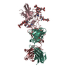
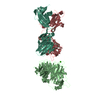
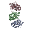
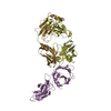
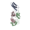
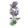
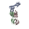
| Entry | Database: PDB / ID: 4mng | ||||||
|---|---|---|---|---|---|---|---|
| Title | Structure of the DP10.7 TCR with CD1d-sulfatide | ||||||
 Components Components |
| ||||||
 Keywords Keywords | IMMUNE SYSTEM / Immunglobulin / Histocompatibility Antigens / CD1 / Immunological / lymphocytes / gamma delta T cell / T cell recognition / self-recognition / myelin / intestinal epithelial lymphocytes / Human / Lipid binding / self-ligand / Glycoproteins / Cell-surface receptors | ||||||
| Function / homology |  Function and homology information Function and homology informationlipid antigen binding / T cell selection / endogenous lipid antigen binding / exogenous lipid antigen binding / antigen processing and presentation, endogenous lipid antigen via MHC class Ib / lipopeptide binding / antigen processing and presentation, exogenous lipid antigen via MHC class Ib / Endosomal/Vacuolar pathway / DAP12 interactions / Antigen Presentation: Folding, assembly and peptide loading of class I MHC ...lipid antigen binding / T cell selection / endogenous lipid antigen binding / exogenous lipid antigen binding / antigen processing and presentation, endogenous lipid antigen via MHC class Ib / lipopeptide binding / antigen processing and presentation, exogenous lipid antigen via MHC class Ib / Endosomal/Vacuolar pathway / DAP12 interactions / Antigen Presentation: Folding, assembly and peptide loading of class I MHC / positive regulation of innate immune response / ER-Phagosome pathway / DAP12 signaling / Immunoregulatory interactions between a Lymphoid and a non-Lymphoid cell / heterotypic cell-cell adhesion / T cell receptor complex / regulation of membrane depolarization / beta-2-microglobulin binding / detection of bacterium / cellular defense response / Neutrophil degranulation / positive regulation of T cell proliferation / cell adhesion molecule binding / regulation of iron ion transport / cellular response to iron(III) ion / antigen processing and presentation of exogenous protein antigen via MHC class Ib, TAP-dependent / negative regulation of iron ion transport / negative regulation of forebrain neuron differentiation / regulation of erythrocyte differentiation / iron ion transport / peptide antigen assembly with MHC class I protein complex / response to molecule of bacterial origin / HFE-transferrin receptor complex / MHC class I peptide loading complex / transferrin transport / negative regulation of receptor-mediated endocytosis / cellular response to iron ion / positive regulation of T cell cytokine production / antigen processing and presentation of endogenous peptide antigen via MHC class I / MHC class I protein complex / peptide antigen assembly with MHC class II protein complex / negative regulation of neurogenesis / multicellular organismal-level iron ion homeostasis / cellular response to nicotine / MHC class II protein complex / positive regulation of receptor-mediated endocytosis / positive regulation of T cell mediated cytotoxicity / negative regulation of epithelial cell proliferation / antigen processing and presentation of exogenous peptide antigen via MHC class II / positive regulation of immune response / peptide antigen binding / phagocytic vesicle membrane / positive regulation of T cell activation / Immunoregulatory interactions between a Lymphoid and a non-Lymphoid cell / sensory perception of smell / positive regulation of cellular senescence / MHC class II protein complex binding / T cell differentiation in thymus / late endosome membrane / negative regulation of neuron projection development / antimicrobial humoral immune response mediated by antimicrobial peptide / antibacterial humoral response / cellular response to lipopolysaccharide / protein refolding / amyloid fibril formation / defense response to Gram-negative bacterium / protein homotetramerization / basolateral plasma membrane / intracellular iron ion homeostasis / adaptive immune response / learning or memory / lysosome / endosome membrane / defense response to Gram-positive bacterium / immune response / external side of plasma membrane / innate immune response / lysosomal membrane / endoplasmic reticulum membrane / structural molecule activity / Golgi apparatus / cell surface / endoplasmic reticulum / protein homodimerization activity / : / identical protein binding / plasma membrane / cytosol / cytoplasm Similarity search - Function | ||||||
| Biological species |   Homo sapiens (human) Homo sapiens (human) | ||||||
| Method |  X-RAY DIFFRACTION / SYNCHROTRON / MOLECULAR REPLACEMENT / Resolution: 3.0058 Å X-RAY DIFFRACTION / SYNCHROTRON / MOLECULAR REPLACEMENT / Resolution: 3.0058 Å | ||||||
 Authors Authors | Luoma, A.M. / Adams, E.J. | ||||||
 Citation Citation | Journal: Immunity / Year: 2013 Title: Crystal Structure of V delta 1 T Cell Receptor in Complex with CD1d-Sulfatide Shows MHC-like Recognition of a Self-Lipid by Human gamma delta T Cells. Authors: Luoma, A.M. / Castro, C.D. / Mayassi, T. / Bembinster, L.A. / Bai, L. / Picard, D. / Anderson, B. / Scharf, L. / Kung, J.E. / Sibener, L.V. / Savage, P.B. / Jabri, B. / Bendelac, A. / Adams, E.J. | ||||||
| History |
|
- Structure visualization
| Structure viewer | Molecule: MolmilJmol/JSmol |
|---|
- Downloads & links
-Download
| PDBx/mmCIF format | 4mng.cif.gz | 256.1 KB | Display | PDBx/mmCIF format |
|---|---|---|---|---|
| PDB format | pdb4mng.ent.gz | 204.6 KB | Display | PDB format |
| PDBx/mmJSON format | 4mng.json.gz | Tree view | PDBx/mmJSON format | |
| Others |  Other downloads Other downloads |
-Validation report
| Arichive directory | https://data.pdbj.org/pub/pdb/validation_reports/mn/4mngftp://data.pdbj.org/pub/pdb/validation_reports/mn/4mng | HTTPS FTP |
|---|
-Related structure data
| Related structure data |  4mnhSC  4mq7SC  4ndmC S: Starting model for refinement C: citing same article ( |
|---|---|
| Similar structure data |


-Links
 PDBj
PDBj


- Assembly
| Deposited unit | 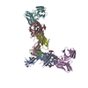
| ||||||||
|---|---|---|---|---|---|---|---|---|---|
| 1 | 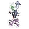
| ||||||||
| 2 | 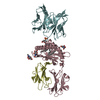
| ||||||||
| Unit cell |
|
-Components
-Protein , 2 types, 4 molecules BDAC
| #1: Protein | Mass: 11660.350 Da / Num. of mol.: 2 / Fragment: Beta-2-microglobulin Source method: isolated from a genetically manipulated source Source: (gene. exp.)  Trichoplusia ni (cabbage looper) / Strain (production host): High Five / References: UniProt: P01887 Trichoplusia ni (cabbage looper) / Strain (production host): High Five / References: UniProt: P01887#2: Protein | Mass: 31972.771 Da / Num. of mol.: 2 / Fragment: CD1d,CD1d Mutation: human CD1d alpha3 domain replaced with mouse CD1d alpha3 domain Source method: isolated from a genetically manipulated source Source: (gene. exp.) Trichoplusia ni (cabbage looper) / Strain (production host): High Five / References: UniProt: P15813, UniProt: Q7TMK5 |
|---|
-Antibody / Sugars , 2 types, 8 molecules EF
| #3: Antibody | Mass: 28534.943 Da / Num. of mol.: 2 Fragment: Single chain of delta and gamma variable domains,Single chain of delta and gamma variable domains Mutation: end of gamma sequence TLVV swapped with KLII Source method: isolated from a genetically manipulated source Source: (gene. exp.) Homo sapiens (human) / Gene: TRA@ / Plasmid: pAK400 / Production host:  #4: Sugar | ChemComp-NAG /  Type: D-saccharide, beta linking / Mass: 221.208 Da / Num. of mol.: 6 Type: D-saccharide, beta linking / Mass: 221.208 Da / Num. of mol.: 6Source method: isolated from a genetically manipulated source Formula: C8H15NO6 |
|---|
-Non-polymers , 2 types, 20 molecules 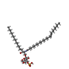

| #5: Chemical |  Mass: 890.301 Da / Num. of mol.: 2 / Source method: obtained synthetically / Formula: C48H91NO11S Mass: 890.301 Da / Num. of mol.: 2 / Source method: obtained synthetically / Formula: C48H91NO11S#6: Water | ChemComp-HOH / | Mass: 18.015 Da / Num. of mol.: 18 / Source method: isolated from a natural source / Formula: H2O |
|---|
-Details
| Has protein modification | Y |
|---|
-Experimental details
-Experiment
| Experiment | Method: X-RAY DIFFRACTION / Number of used crystals: 1 |
|---|
- Sample preparation
| Crystal | Density Matthews: 5.54 Å3/Da / Density % sol: 77.79 % |
|---|---|
| Crystal grow | Temperature: 292 K / Method: vapor diffusion, sitting drop Details: 11% PEG 4000 5% isopropanol 2 mM glutathione reduced/oxidized 50 mM tri-sodium citrate, VAPOR DIFFUSION, SITTING DROP, temperature 292K |
-Data collection
| Diffraction | Mean temperature: 100 K |
|---|---|
| Diffraction source | Source: SYNCHROTRON / Site: APS  / Beamline: 23-ID-D / Wavelength: 1.03321 Å / Beamline: 23-ID-D / Wavelength: 1.03321 Å |
| Detector | Type: MAR scanner 300 mm plate / Detector: IMAGE PLATE / Date: Mar 24, 2013 |
| Radiation | Monochromator: Si(111) double crystal monochromator / Protocol: SINGLE WAVELENGTH / Monochromatic (M) / Laue (L): M / Scattering type: x-ray |
| Radiation wavelength | Wavelength: 1.03321 Å / Relative weight: 1 |
| Reflection | Resolution: 3→50 Å / Num. all: 61100 / Num. obs: 61100 / % possible obs: 99.2 % / Observed criterion σ(F): 0 / Observed criterion σ(I): -3 / Redundancy: 4 % |
| Reflection shell | Resolution: 3→3.05 Å / % possible all: 95.7 |
- Processing
| Software |
| |||||||||||||||||||||||||||||||||||||||||||||||||||||||||||||||||||||||||||||||||||||||||||||||||||||||||||||||||||||||||||||||||||||||||||||||||||||||||||||||||
|---|---|---|---|---|---|---|---|---|---|---|---|---|---|---|---|---|---|---|---|---|---|---|---|---|---|---|---|---|---|---|---|---|---|---|---|---|---|---|---|---|---|---|---|---|---|---|---|---|---|---|---|---|---|---|---|---|---|---|---|---|---|---|---|---|---|---|---|---|---|---|---|---|---|---|---|---|---|---|---|---|---|---|---|---|---|---|---|---|---|---|---|---|---|---|---|---|---|---|---|---|---|---|---|---|---|---|---|---|---|---|---|---|---|---|---|---|---|---|---|---|---|---|---|---|---|---|---|---|---|---|---|---|---|---|---|---|---|---|---|---|---|---|---|---|---|---|---|---|---|---|---|---|---|---|---|---|---|---|---|---|---|---|
| Refinement | Method to determine structure: MOLECULAR REPLACEMENT Starting model: PDB ENTRY 4MNH and 4MQ7 Resolution: 3.0058→49.652 Å / SU ML: 0.35 / σ(F): 1.97 / Phase error: 21.47 / Stereochemistry target values: ML
| |||||||||||||||||||||||||||||||||||||||||||||||||||||||||||||||||||||||||||||||||||||||||||||||||||||||||||||||||||||||||||||||||||||||||||||||||||||||||||||||||
| Solvent computation | Shrinkage radii: 0.9 Å / VDW probe radii: 1.11 Å / Solvent model: FLAT BULK SOLVENT MODEL | |||||||||||||||||||||||||||||||||||||||||||||||||||||||||||||||||||||||||||||||||||||||||||||||||||||||||||||||||||||||||||||||||||||||||||||||||||||||||||||||||
| Refinement step | Cycle: LAST / Resolution: 3.0058→49.652 Å
| |||||||||||||||||||||||||||||||||||||||||||||||||||||||||||||||||||||||||||||||||||||||||||||||||||||||||||||||||||||||||||||||||||||||||||||||||||||||||||||||||
| Refine LS restraints |
| |||||||||||||||||||||||||||||||||||||||||||||||||||||||||||||||||||||||||||||||||||||||||||||||||||||||||||||||||||||||||||||||||||||||||||||||||||||||||||||||||
| LS refinement shell |
|
