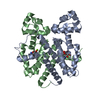
 Movie
Movie Controller


+ Open data

- Basic information
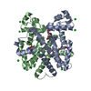
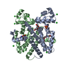
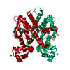
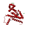
| Entry | Database: PDB / ID: 6rbl | ||||||
|---|---|---|---|---|---|---|---|
| Title | TETR(D) E147A MUTANT IN COMPLEX WITH DOXYCYCLINE AND MAGNESIUM | ||||||
 Components Components | Tetracycline repressor protein class D | ||||||
 Keywords Keywords | TRANSCRIPTION / TRANSCRIPTION REGULATION | ||||||
| Function / homology |  Function and homology information Function and homology informationtranscription cis-regulatory region binding / DNA-binding transcription factor activity / response to antibiotic / negative regulation of DNA-templated transcription / DNA-templated transcription / metal ion binding Similarity search - Function | ||||||
| Biological species |  | ||||||
| Method |  X-RAY DIFFRACTION / MOLECULAR REPLACEMENT / Resolution: 1.9 Å X-RAY DIFFRACTION / MOLECULAR REPLACEMENT / Resolution: 1.9 Å | ||||||
 Authors Authors | Hinrichs, W. / Palm, G.J. / Berndt, L. / Girbardt, B. | ||||||
 Citation Citation | Journal: Biochim Biophys Acta Proteins Proteom / Year: 2020 Title: Thermodynamics, cooperativity and stability of the tetracycline repressor (TetR) upon tetracycline binding. Authors: Palm, G.J. / Buchholz, I. / Werten, S. / Girbardt, B. / Berndt, L. / Delcea, M. / Hinrichs, W. #1: Journal: FEBS J. / Year: 2016Title: Modular organisation of inducer recognition and allostery in the tetracycline repressor. Authors: Werten, S. / Schneider, J. / Palm, G.J. / Hinrichs, W. | ||||||
| History |
|
- Structure visualization
| Structure viewer | Molecule: MolmilJmol/JSmol |
|---|
- Downloads & links
-Download
| PDBx/mmCIF format | 6rbl.cif.gz | 104.4 KB | Display | PDBx/mmCIF format |
|---|---|---|---|---|
| PDB format | pdb6rbl.ent.gz | 80 KB | Display | PDB format |
| PDBx/mmJSON format | 6rbl.json.gz | Tree view | PDBx/mmJSON format | |
| Others |  Other downloads Other downloads |
-Validation report
| Arichive directory | https://data.pdbj.org/pub/pdb/validation_reports/rb/6rblftp://data.pdbj.org/pub/pdb/validation_reports/rb/6rbl | HTTPS FTP |
|---|
-Related structure data
| Related structure data |  2xrlC  4b3aC  6fplSC  6fpmC  6ftsC  6qjwC  6qjxC  6rbmC  6rcrC  6rgxC S: Starting model for refinement C: citing same article ( |
|---|---|
| Similar structure data |


-Links
 PDBj
PDBj- Assembly
| Deposited unit | 
| |||||||||
|---|---|---|---|---|---|---|---|---|---|---|
| 1 | 
| |||||||||
| Unit cell |
| |||||||||
| Components on special symmetry positions |
|
-Components
| #1: Protein | Mass: 23230.299 Da / Num. of mol.: 1 / Mutation: A2S, E147A Source method: isolated from a genetically manipulated source Details: RA1-plasmid / Source: (gene. exp.) | ||
|---|---|---|---|
| #2: Chemical | ChemComp-DXT / (  Mass: 444.435 Da / Num. of mol.: 1 / Source method: obtained synthetically / Formula: C22H24N2O8 / Feature type: SUBJECT OF INVESTIGATION / Comment: antibiotic*YM Mass: 444.435 Da / Num. of mol.: 1 / Source method: obtained synthetically / Formula: C22H24N2O8 / Feature type: SUBJECT OF INVESTIGATION / Comment: antibiotic*YM | ||
| #3: Chemical | ChemComp-MG /   Mass: 24.305 Da / Num. of mol.: 1 / Source method: obtained synthetically / Formula: Mg Mass: 24.305 Da / Num. of mol.: 1 / Source method: obtained synthetically / Formula: Mg | ||
| #4: Chemical | ChemComp-CL /   Mass: 35.453 Da / Num. of mol.: 6 / Source method: obtained synthetically / Formula: Cl Mass: 35.453 Da / Num. of mol.: 6 / Source method: obtained synthetically / Formula: Cl#5: Water | ChemComp-HOH / |  Mass: 18.015 Da / Num. of mol.: 161 / Source method: isolated from a natural source / Formula: H2O Mass: 18.015 Da / Num. of mol.: 161 / Source method: isolated from a natural source / Formula: H2O |
-Experimental details
-Experiment
| Experiment | Method: X-RAY DIFFRACTION / Number of used crystals: 1 |
|---|
- Sample preparation
| Crystal | Density Matthews: 2.25 Å3/Da / Density % sol: 45.37 % |
|---|---|
| Crystal grow | Temperature: 295 K / Method: vapor diffusion, hanging drop Details: Precipitant: 2.0M (NH4)2SO4, 3% PEG 400, 0.1M MES pH 6.5, Protein: 0.1mL protein, 0.1,L 2mM doxyTc, 0.001mL 3M MgCl2, 100mM NaCl, 50mM Tris pH8.0, 1/3 protein/precipitant |
-Data collection
| Diffraction | Mean temperature: 100 K / Serial crystal experiment: N |
|---|---|
| Diffraction source | Source: ROTATING ANODE / Type: RIGAKU MICROMAX-007 / Wavelength: 1.5418 Å |
| Detector | Type: RIGAKU SATURN 70 / Detector: CCD / Date: Apr 18, 2007 / Details: mirror |
| Radiation | Monochromator: Mirror / Protocol: SINGLE WAVELENGTH / Monochromatic (M) / Laue (L): M / Scattering type: x-ray |
| Radiation wavelength | Wavelength: 1.5418 Å / Relative weight: 1 |
| Reflection | Resolution: 1.9→34.17 Å / Num. obs: 17126 / % possible obs: 99.4 % / Redundancy: 7.1 % / Biso Wilson estimate: 21 Å2 / Rmerge(I) obs: 0.099 / Net I/σ(I): 12.3 |
| Reflection shell | Resolution: 1.9→1.97 Å / Redundancy: 4.29 % / Rmerge(I) obs: 0.371 / Mean I/σ(I) obs: 1.9 / Num. unique obs: 1617 / % possible all: 98.2 |
- Processing
| Software |
| ||||||||||||||||||||||||||||||||||||||||||||||||||||||||||||||||||||||||||||||||||||||||||||||||||||||||||||||||||||||||||||||||||||||||||||||||||||||||||||||||||||||||||||||||||||||
|---|---|---|---|---|---|---|---|---|---|---|---|---|---|---|---|---|---|---|---|---|---|---|---|---|---|---|---|---|---|---|---|---|---|---|---|---|---|---|---|---|---|---|---|---|---|---|---|---|---|---|---|---|---|---|---|---|---|---|---|---|---|---|---|---|---|---|---|---|---|---|---|---|---|---|---|---|---|---|---|---|---|---|---|---|---|---|---|---|---|---|---|---|---|---|---|---|---|---|---|---|---|---|---|---|---|---|---|---|---|---|---|---|---|---|---|---|---|---|---|---|---|---|---|---|---|---|---|---|---|---|---|---|---|---|---|---|---|---|---|---|---|---|---|---|---|---|---|---|---|---|---|---|---|---|---|---|---|---|---|---|---|---|---|---|---|---|---|---|---|---|---|---|---|---|---|---|---|---|---|---|---|---|---|
| Refinement | Method to determine structure: MOLECULAR REPLACEMENT Starting model: 6FPL Resolution: 1.9→34.17 Å / Cor.coef. Fo:Fc: 0.945 / Cor.coef. Fo:Fc free: 0.926 / SU B: 9.102 / SU ML: 0.117 / Cross valid method: THROUGHOUT / ESU R: 0.196 / ESU R Free: 0.167 / Details: HYDROGENS HAVE BEEN ADDED IN THE RIDING POSITIONS
| ||||||||||||||||||||||||||||||||||||||||||||||||||||||||||||||||||||||||||||||||||||||||||||||||||||||||||||||||||||||||||||||||||||||||||||||||||||||||||||||||||||||||||||||||||||||
| Solvent computation | Ion probe radii: 0.8 Å / Shrinkage radii: 0.8 Å / VDW probe radii: 1.2 Å | ||||||||||||||||||||||||||||||||||||||||||||||||||||||||||||||||||||||||||||||||||||||||||||||||||||||||||||||||||||||||||||||||||||||||||||||||||||||||||||||||||||||||||||||||||||||
| Displacement parameters | Biso mean: 21.97 Å2
| ||||||||||||||||||||||||||||||||||||||||||||||||||||||||||||||||||||||||||||||||||||||||||||||||||||||||||||||||||||||||||||||||||||||||||||||||||||||||||||||||||||||||||||||||||||||
| Refinement step | Cycle: 1 / Resolution: 1.9→34.17 Å
| ||||||||||||||||||||||||||||||||||||||||||||||||||||||||||||||||||||||||||||||||||||||||||||||||||||||||||||||||||||||||||||||||||||||||||||||||||||||||||||||||||||||||||||||||||||||
| Refine LS restraints |
|