 Movie
Movie Controller

[English] 日本語
 Yorodumi
- PDB-2fj1: Crystal Structure Analysis of Tet Repressor (class D) in Complex ... -
+ Open data

- Basic information
| Entry | Database: PDB / ID: 2fj1 | ||||||
|---|---|---|---|---|---|---|---|
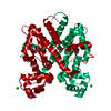
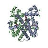
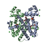
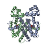
| Title | Crystal Structure Analysis of Tet Repressor (class D) in Complex with 7-Chlortetracycline-Nickel(II) | ||||||
 Components Components | Tetracycline repressor protein class D | ||||||
 Keywords Keywords | TRANSCRIPTION REGULATOR / TRANSCRIPTION REGULATION / helix-turn-helix / metal coordination | ||||||
| Function / homology |  Function and homology information Function and homology informationtranscription cis-regulatory region binding / DNA-binding transcription factor activity / response to antibiotic / negative regulation of DNA-templated transcription / metal ion binding Similarity search - Function | ||||||
| Biological species |  | ||||||
| Method |  X-RAY DIFFRACTION / MOLECULAR REPLACEMENT / Resolution: 2.2 Å X-RAY DIFFRACTION / MOLECULAR REPLACEMENT / Resolution: 2.2 Å | ||||||
 Authors Authors | Orth, P. / Saenger, W. / Palm, G.J. / Hinrichs, W. | ||||||
 Citation Citation | Journal: J.Biol.Inorg.Chem. / Year: 2008 Title: Specific binding of divalent metal ions to tetracycline and to the Tet repressor/tetracycline complex. Authors: Palm, G.J. / Lederer, T. / Orth, P. / Saenger, W. / Takahashi, M. / Hillen, W. / Hinrichs, W. #1: Journal: Science / Year: 1994Title: Structure of the TET repressor-tetracycline complex and regulation of antibiotic resistance Authors: Hinrichs, W. / Kisker, C. / Duevel, M. / Mueller, A. / Tovar, K. / Hillen, W. / Saenger, W. | ||||||
| History |
|
- Structure visualization
| Structure viewer | Molecule: MolmilJmol/JSmol |
|---|
- Downloads & links
-Download
| PDBx/mmCIF format | 2fj1.cif.gz | 59.8 KB | Display | PDBx/mmCIF format |
|---|---|---|---|---|
| PDB format | pdb2fj1.ent.gz | 43 KB | Display | PDB format |
| PDBx/mmJSON format | 2fj1.json.gz | Tree view | PDBx/mmJSON format | |
| Others |  Other downloads Other downloads |
-Validation report
| Arichive directory | https://data.pdbj.org/pub/pdb/validation_reports/fj/2fj1ftp://data.pdbj.org/pub/pdb/validation_reports/fj/2fj1 | HTTPS FTP |
|---|
-Related structure data
| Related structure data |  2vkeC  2tctS S: Starting model for refinement C: citing same article ( |
|---|---|
| Similar structure data |


-Links
 PDBj
PDBj- Assembly
| Deposited unit | 
| ||||||||
|---|---|---|---|---|---|---|---|---|---|
| 1 | 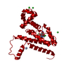
| ||||||||
| Unit cell |
|
-Components
| #1: Protein | Mass: 23288.334 Da / Num. of mol.: 1 / Fragment: residues 2-208 / Mutation: A2S Source method: isolated from a genetically manipulated source Source: (gene. exp.) | ||||
|---|---|---|---|---|---|
| #2: Chemical | ChemComp-NI /   Mass: 58.693 Da / Num. of mol.: 1 / Source method: obtained synthetically / Formula: Ni Mass: 58.693 Da / Num. of mol.: 1 / Source method: obtained synthetically / Formula: Ni | ||||
| #3: Chemical |   Mass: 35.453 Da / Num. of mol.: 3 / Source method: obtained synthetically / Formula: Cl Mass: 35.453 Da / Num. of mol.: 3 / Source method: obtained synthetically / Formula: Cl#4: Chemical | ChemComp-CTC / |   Mass: 478.880 Da / Num. of mol.: 1 / Source method: obtained synthetically / Formula: C22H23ClN2O8 Mass: 478.880 Da / Num. of mol.: 1 / Source method: obtained synthetically / Formula: C22H23ClN2O8#5: Water | ChemComp-HOH / |  Mass: 18.015 Da / Num. of mol.: 102 / Source method: isolated from a natural source / Formula: H2O Mass: 18.015 Da / Num. of mol.: 102 / Source method: isolated from a natural source / Formula: H2O |
-Experimental details
-Experiment
| Experiment | Method: X-RAY DIFFRACTION / Number of used crystals: 1 |
|---|
- Sample preparation
| Crystal | Density Matthews: 2.31 Å3/Da / Density % sol: 46.67 % |
|---|---|
| Crystal grow | Temperature: 293 K / Method: vapor diffusion, hanging drop / pH: 7.2 Details: 1.58 M Ammonium sulphate, pH 7.2, VAPOR DIFFUSION, HANGING DROP, temperature 293.0K |
-Data collection
| Diffraction | Mean temperature: 277 K |
|---|---|
| Diffraction source | Source: ROTATING ANODE / Type: ENRAF-NONIUS FR571 / Wavelength: 1.5418 Å |
| Detector | Type: MARRESEARCH / Detector: IMAGE PLATE / Date: Jan 5, 1997 |
| Radiation | Monochromator: graphite / Protocol: SINGLE WAVELENGTH / Monochromatic (M) / Laue (L): M / Scattering type: x-ray |
| Radiation wavelength | Wavelength: 1.5418 Å / Relative weight: 1 |
| Reflection | Resolution: 2.2→64.5 Å / Num. all: 10706 / Num. obs: 10706 / % possible obs: 97.6 % / Observed criterion σ(F): 0 / Observed criterion σ(I): 0 / Redundancy: 4.5 % / Biso Wilson estimate: 32.1 Å2 / Rsym value: 0.06 / Net I/σ(I): 16.4 |
| Reflection shell | Resolution: 2.2→2.258 Å / Mean I/σ(I) obs: 3.2 / Num. unique all: 751 / Rsym value: 0.268 |
- Processing
| Software |
| ||||||||||||||||||||||||||||||||||||||||||||||||||||||||||||||||||||||||||||||||||||||||||||||||||||
|---|---|---|---|---|---|---|---|---|---|---|---|---|---|---|---|---|---|---|---|---|---|---|---|---|---|---|---|---|---|---|---|---|---|---|---|---|---|---|---|---|---|---|---|---|---|---|---|---|---|---|---|---|---|---|---|---|---|---|---|---|---|---|---|---|---|---|---|---|---|---|---|---|---|---|---|---|---|---|---|---|---|---|---|---|---|---|---|---|---|---|---|---|---|---|---|---|---|---|---|---|---|
| Refinement | Method to determine structure: MOLECULAR REPLACEMENT Starting model: PDB ENTRY 2TCT Resolution: 2.2→64.55 Å / Cor.coef. Fo:Fc: 0.972 / Cor.coef. Fo:Fc free: 0.942 / SU B: 5.467 / SU ML: 0.133 / Cross valid method: THROUGHOUT / σ(F): 0 / σ(I): 0 / ESU R: 0.24 / ESU R Free: 0.2 / Stereochemistry target values: MAXIMUM LIKELIHOOD / Details: HYDROGENS HAVE BEEN ADDED IN THE RIDING POSITIONS
| ||||||||||||||||||||||||||||||||||||||||||||||||||||||||||||||||||||||||||||||||||||||||||||||||||||
| Solvent computation | Ion probe radii: 0.8 Å / Shrinkage radii: 0.8 Å / VDW probe radii: 1.4 Å / Solvent model: BABINET MODEL WITH MASK | ||||||||||||||||||||||||||||||||||||||||||||||||||||||||||||||||||||||||||||||||||||||||||||||||||||
| Displacement parameters | Biso mean: 33.999 Å2
| ||||||||||||||||||||||||||||||||||||||||||||||||||||||||||||||||||||||||||||||||||||||||||||||||||||
| Refinement step | Cycle: LAST / Resolution: 2.2→64.55 Å
| ||||||||||||||||||||||||||||||||||||||||||||||||||||||||||||||||||||||||||||||||||||||||||||||||||||
| Refine LS restraints |
| ||||||||||||||||||||||||||||||||||||||||||||||||||||||||||||||||||||||||||||||||||||||||||||||||||||
| LS refinement shell | Resolution: 2.2→2.258 Å / Total num. of bins used: 20 /
|
