 Movie
Movie Controller

[English] 日本語
 Yorodumi
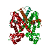
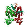
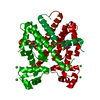
- PDB-4b3a: Tetracycline repressor class D mutant H100A in complex with tetra... -
+ Open data

- Basic information
| Entry | Database: PDB / ID: 4b3a | ||||||
|---|---|---|---|---|---|---|---|
| Title | Tetracycline repressor class D mutant H100A in complex with tetracycline | ||||||
 Components Components | TETRACYCLINE REPRESSOR PROTEIN CLASS D | ||||||
 Keywords Keywords | TRANSCRIPTION / TET-REPRESSOR | ||||||
| Function / homology |  Function and homology information Function and homology informationtranscription cis-regulatory region binding / DNA-binding transcription factor activity / response to antibiotic / negative regulation of DNA-templated transcription / DNA-templated transcription / metal ion binding Similarity search - Function | ||||||
| Biological species |  | ||||||
| Method |  X-RAY DIFFRACTION / MOLECULAR REPLACEMENT / Resolution: 1.7 Å X-RAY DIFFRACTION / MOLECULAR REPLACEMENT / Resolution: 1.7 Å | ||||||
 Authors Authors | Eltschkner, S. / Schindler, S. / Palm, G.J. / Schneider, J. / Hinrichs, W. | ||||||
 Citation Citation | Journal: Biochim Biophys Acta Proteins Proteom / Year: 2020 Title: Thermodynamics, cooperativity and stability of the tetracycline repressor (TetR) upon tetracycline binding. Authors: Palm, G.J. / Buchholz, I. / Werten, S. / Girbardt, B. / Berndt, L. / Delcea, M. / Hinrichs, W. | ||||||
| History |
|
- Structure visualization
| Structure viewer | Molecule: MolmilJmol/JSmol |
|---|
- Downloads & links
-Download
| PDBx/mmCIF format | 4b3a.cif.gz | 100.8 KB | Display | PDBx/mmCIF format |
|---|---|---|---|---|
| PDB format | pdb4b3a.ent.gz | 77.6 KB | Display | PDB format |
| PDBx/mmJSON format | 4b3a.json.gz | Tree view | PDBx/mmJSON format | |
| Others |  Other downloads Other downloads |
-Validation report
| Arichive directory | https://data.pdbj.org/pub/pdb/validation_reports/b3/4b3aftp://data.pdbj.org/pub/pdb/validation_reports/b3/4b3a | HTTPS FTP |
|---|
-Related structure data
| Related structure data |  2xrlC  6fplC  6fpmC  6ftsC  6qjwC  6qjxC  6rblC  6rbmC  6rcrC  6rgxC  2vkeS S: Starting model for refinement C: citing same article ( |
|---|---|
| Similar structure data |


-Links
 PDBj
PDBj
- Assembly
| Deposited unit | 
| ||||||||||||
|---|---|---|---|---|---|---|---|---|---|---|---|---|---|
| 1 | 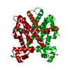
| ||||||||||||
| Unit cell |
| ||||||||||||
| Components on special symmetry positions |
|
-Components
| #1: Protein | Mass: 23221.264 Da / Num. of mol.: 1 / Fragment: RESIDUES 2-208 / Mutation: YES Source method: isolated from a genetically manipulated source Source: (gene. exp.) | ||||||
|---|---|---|---|---|---|---|---|
| #2: Chemical | ChemComp-TAC / 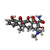  Mass: 444.435 Da / Num. of mol.: 1 / Source method: obtained synthetically / Formula: C22H24N2O8 / Comment: medication, antibiotic*YM Mass: 444.435 Da / Num. of mol.: 1 / Source method: obtained synthetically / Formula: C22H24N2O8 / Comment: medication, antibiotic*YM | ||||||
| #3: Chemical |   Mass: 35.453 Da / Num. of mol.: 3 / Source method: obtained synthetically / Formula: Cl Mass: 35.453 Da / Num. of mol.: 3 / Source method: obtained synthetically / Formula: Cl#4: Chemical | ChemComp-MG / |   Mass: 24.305 Da / Num. of mol.: 1 / Source method: obtained synthetically / Formula: Mg Mass: 24.305 Da / Num. of mol.: 1 / Source method: obtained synthetically / Formula: Mg#5: Water | ChemComp-HOH / |  Mass: 18.015 Da / Num. of mol.: 130 / Source method: isolated from a natural source / Formula: H2O Mass: 18.015 Da / Num. of mol.: 130 / Source method: isolated from a natural source / Formula: H2OSequence details | N-TERMINAL MET REMOVED. RESIDUES 209 - 218 REMOVED BY CLONING. | |
-Experimental details
-Experiment
| Experiment | Method: X-RAY DIFFRACTION / Number of used crystals: 1 |
|---|
- Sample preparation
| Crystal | Density Matthews: 2.2 Å3/Da / Density % sol: 44 % / Description: NONE |
|---|---|
| Crystal grow | pH: 6.5 Details: 0.5 M AMMONIUM SULPHATE, 1 M LITHIUM SULPHATE, 0.1 M MES PH 6.5 |
-Data collection
| Diffraction | Mean temperature: 100 K |
|---|---|
| Diffraction source | Source: ROTATING ANODE / Type: RIGAKU MICROMAX-007 / Wavelength: 1.5418 |
| Detector | Type: RIGAKU CCD / Detector: CCD / Date: Apr 17, 2007 / Details: OSMIC MULTILAYER |
| Radiation | Monochromator: OSMIC MULTILAYER / Protocol: SINGLE WAVELENGTH / Monochromatic (M) / Laue (L): M / Scattering type: x-ray |
| Radiation wavelength | Wavelength: 1.5418 Å / Relative weight: 1 |
| Reflection | Resolution: 1.7→29.79 Å / Num. obs: 19362 / % possible obs: 82.6 % / Observed criterion σ(I): -3 / Redundancy: 5.86 % / Rmerge(I) obs: 0.05 / Net I/σ(I): 20.4 |
| Reflection shell | Resolution: 1.7→1.76 Å / Redundancy: 1.15 % / Rmerge(I) obs: 0.46 / Mean I/σ(I) obs: 0.9 / % possible all: 17.4 |
- Processing
| Software |
| ||||||||||||||||||||||||||||||||||||||||||||||||||||||||||||||||||||||||||||||||||||||||||||||||||||||||||||||||||||||||||||||||||||||||||||||||||||||||||||||||||||||||||||||||||||||
|---|---|---|---|---|---|---|---|---|---|---|---|---|---|---|---|---|---|---|---|---|---|---|---|---|---|---|---|---|---|---|---|---|---|---|---|---|---|---|---|---|---|---|---|---|---|---|---|---|---|---|---|---|---|---|---|---|---|---|---|---|---|---|---|---|---|---|---|---|---|---|---|---|---|---|---|---|---|---|---|---|---|---|---|---|---|---|---|---|---|---|---|---|---|---|---|---|---|---|---|---|---|---|---|---|---|---|---|---|---|---|---|---|---|---|---|---|---|---|---|---|---|---|---|---|---|---|---|---|---|---|---|---|---|---|---|---|---|---|---|---|---|---|---|---|---|---|---|---|---|---|---|---|---|---|---|---|---|---|---|---|---|---|---|---|---|---|---|---|---|---|---|---|---|---|---|---|---|---|---|---|---|---|---|
| Refinement | Method to determine structure: MOLECULAR REPLACEMENT Starting model: PDB ENTRY 2VKE Resolution: 1.7→29.8 Å / Cor.coef. Fo:Fc: 0.967 / Cor.coef. Fo:Fc free: 0.952 / SU B: 8.479 / SU ML: 0.116 / Cross valid method: THROUGHOUT / ESU R: 0.148 / ESU R Free: 0.143 / Stereochemistry target values: MAXIMUM LIKELIHOOD Details: HYDROGENS HAVE BEEN ADDED IN THE RIDING POSITIONS. U VALUES WITH TLS ADDED
| ||||||||||||||||||||||||||||||||||||||||||||||||||||||||||||||||||||||||||||||||||||||||||||||||||||||||||||||||||||||||||||||||||||||||||||||||||||||||||||||||||||||||||||||||||||||
| Solvent computation | Ion probe radii: 0.8 Å / Shrinkage radii: 0.8 Å / VDW probe radii: 1.2 Å / Solvent model: MASK | ||||||||||||||||||||||||||||||||||||||||||||||||||||||||||||||||||||||||||||||||||||||||||||||||||||||||||||||||||||||||||||||||||||||||||||||||||||||||||||||||||||||||||||||||||||||
| Displacement parameters | Biso mean: 42.319 Å2
| ||||||||||||||||||||||||||||||||||||||||||||||||||||||||||||||||||||||||||||||||||||||||||||||||||||||||||||||||||||||||||||||||||||||||||||||||||||||||||||||||||||||||||||||||||||||
| Refinement step | Cycle: LAST / Resolution: 1.7→29.8 Å
| ||||||||||||||||||||||||||||||||||||||||||||||||||||||||||||||||||||||||||||||||||||||||||||||||||||||||||||||||||||||||||||||||||||||||||||||||||||||||||||||||||||||||||||||||||||||
| Refine LS restraints |
|