Movie
Movie Controller


+ Open data

- Basic information
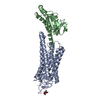
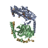
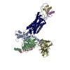
| Entry | Database: PDB / ID: 6qno | ||||||||||||||||||
|---|---|---|---|---|---|---|---|---|---|---|---|---|---|---|---|---|---|---|---|
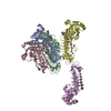
| Title | Rhodopsin-Gi protein complex | ||||||||||||||||||
 Components Components |
| ||||||||||||||||||
 Keywords Keywords | SIGNALING PROTEIN / GPCR and G protein complex | ||||||||||||||||||
| Function / homology |  Function and homology information Function and homology informationOpsins / VxPx cargo-targeting to cilium / sperm head plasma membrane / transducin-mediated opsin signaling pathway / opsin binding / The canonical retinoid cycle in rods (twilight vision) / absorption of visible light / G protein-coupled opsin signaling pathway / Olfactory Signaling Pathway / 11-cis retinal binding ...Opsins / VxPx cargo-targeting to cilium / sperm head plasma membrane / transducin-mediated opsin signaling pathway / opsin binding / The canonical retinoid cycle in rods (twilight vision) / absorption of visible light / G protein-coupled opsin signaling pathway / Olfactory Signaling Pathway / 11-cis retinal binding / Sensory perception of sweet, bitter, and umami (glutamate) taste / Synthesis, secretion, and inactivation of Glucagon-like Peptide-1 (GLP-1) / G protein-coupled photoreceptor activity / photoreceptor inner segment membrane / cellular response to light stimulus / G protein-coupled receptor complex / Inactivation, recovery and regulation of the phototransduction cascade / Activation of the phototransduction cascade / outer membrane / arrestin family protein binding / Activation of G protein gated Potassium channels / G-protein activation / G beta:gamma signalling through PI3Kgamma / Prostacyclin signalling through prostacyclin receptor / G beta:gamma signalling through PLC beta / ADP signalling through P2Y purinoceptor 1 / Thromboxane signalling through TP receptor / Presynaptic function of Kainate receptors / G beta:gamma signalling through CDC42 / Inhibition of voltage gated Ca2+ channels via Gbeta/gamma subunits / G alpha (12/13) signalling events / Glucagon-type ligand receptors / G beta:gamma signalling through BTK / ADP signalling through P2Y purinoceptor 12 / Adrenaline,noradrenaline inhibits insulin secretion / Cooperation of PDCL (PhLP1) and TRiC/CCT in G-protein beta folding / Ca2+ pathway / G alpha (z) signalling events / Thrombin signalling through proteinase activated receptors (PARs) / Extra-nuclear estrogen signaling / G alpha (s) signalling events / G alpha (q) signalling events / photoreceptor outer segment membrane / Glucagon-like Peptide-1 (GLP1) regulates insulin secretion / G alpha (i) signalling events / High laminar flow shear stress activates signaling by PIEZO1 and PECAM1:CDH5:KDR in endothelial cells / Vasopressin regulates renal water homeostasis via Aquaporins / phototransduction, visible light / phototransduction / response to light stimulus / photoreceptor outer segment / G-protein alpha-subunit binding / regulation of eating behavior / adenylate cyclase inhibitor activity / positive regulation of protein localization to cell cortex / T cell migration / positive regulation of relaxation of smooth muscle / Adenylate cyclase inhibitory pathway / visual perception / D2 dopamine receptor binding / guanyl-nucleotide exchange factor activity / adenylate cyclase-inhibiting serotonin receptor signaling pathway / G protein-coupled serotonin receptor binding / cellular response to forskolin / mast cell degranulation / regulation of mitotic spindle organization / chemokine-mediated signaling pathway / Regulation of insulin secretion / neuropeptide signaling pathway / response to prostaglandin E / positive regulation of cholesterol biosynthetic process / G protein-coupled receptor binding / response to peptide hormone / G-protein beta/gamma-subunit complex binding / adenylate cyclase-modulating G protein-coupled receptor signaling pathway / adenylate cyclase-inhibiting G protein-coupled receptor signaling pathway / cell-cell junction / photoreceptor disc membrane / GDP binding / ADP signalling through P2Y purinoceptor 12 / Adrenaline,noradrenaline inhibits insulin secretion / G alpha (z) signalling events / cellular response to catecholamine stimulus / ADORA2B mediated anti-inflammatory cytokines production / adenylate cyclase-activating dopamine receptor signaling pathway / GPER1 signaling / sperm midpiece / cellular response to prostaglandin E stimulus / heterotrimeric G-protein complex / G-protein beta-subunit binding / sensory perception of taste / signaling receptor complex adaptor activity / adenylate cyclase-activating G protein-coupled receptor signaling pathway / retina development in camera-type eye / GTPase binding / G protein activity / midbody / cell cortex / G alpha (i) signalling events / G alpha (s) signalling events Similarity search - Function | ||||||||||||||||||
| Biological species |  Homo sapiens (human) Homo sapiens (human) | ||||||||||||||||||
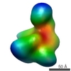
| Method | ELECTRON MICROSCOPY / single particle reconstruction / cryo EM / Resolution: 4.38 Å | ||||||||||||||||||
 Authors Authors | Tsai, C.-J. / Marino, J. / Adaixo, R.J. / Pamula, F. / Muehle, J. / Maeda, S. / Flock, T. / Taylor, N.M.I. / Mohammed, I. / Matile, H. ...Tsai, C.-J. / Marino, J. / Adaixo, R.J. / Pamula, F. / Muehle, J. / Maeda, S. / Flock, T. / Taylor, N.M.I. / Mohammed, I. / Matile, H. / Dawson, R.J.P. / Deupi, X. / Stahlberg, H. / Schertler, G.F.X. | ||||||||||||||||||
| Funding support |  Switzerland, 5items Switzerland, 5items
| ||||||||||||||||||
 Citation Citation | Journal: Elife / Year: 2019 Title: Cryo-EM structure of the rhodopsin-Gαi-βγ complex reveals binding of the rhodopsin C-terminal tail to the gβ subunit. Authors: Ching-Ju Tsai / Jacopo Marino / Ricardo Adaixo / Filip Pamula / Jonas Muehle / Shoji Maeda / Tilman Flock / Nicholas Mi Taylor / Inayatulla Mohammed / Hugues Matile / Roger Jp Dawson / ...Authors: Ching-Ju Tsai / Jacopo Marino / Ricardo Adaixo / Filip Pamula / Jonas Muehle / Shoji Maeda / Tilman Flock / Nicholas Mi Taylor / Inayatulla Mohammed / Hugues Matile / Roger Jp Dawson / Xavier Deupi / Henning Stahlberg / Gebhard Schertler / Abstract: One of the largest membrane protein families in eukaryotes are G protein-coupled receptors (GPCRs). GPCRs modulate cell physiology by activating diverse intracellular transducers, prominently ...One of the largest membrane protein families in eukaryotes are G protein-coupled receptors (GPCRs). GPCRs modulate cell physiology by activating diverse intracellular transducers, prominently heterotrimeric G proteins. The recent surge in structural data has expanded our understanding of GPCR-mediated signal transduction. However, many aspects, including the existence of transient interactions, remain elusive. We present the cryo-EM structure of the light-sensitive GPCR rhodopsin in complex with heterotrimeric Gi. Our density map reveals the receptor C-terminal tail bound to the Gβ subunit of the G protein, providing a structural foundation for the role of the C-terminal tail in GPCR signaling, and of Gβ as scaffold for recruiting Gα subunits and G protein-receptor kinases. By comparing available complexes, we found a small set of common anchoring points that are G protein-subtype specific. Taken together, our structure and analysis provide new structural basis for the molecular events of the GPCR signaling pathway. | ||||||||||||||||||
| History |
|
- Structure visualization
| Movie |
Movie viewer |
|---|---|
| Structure viewer | Molecule: MolmilJmol/JSmol |
- Downloads & links
-Download
| PDBx/mmCIF format | 6qno.cif.gz | 252.4 KB | Display | PDBx/mmCIF format |
|---|---|---|---|---|
| PDB format | pdb6qno.ent.gz | 194.7 KB | Display | PDB format |
| PDBx/mmJSON format | 6qno.json.gz | Tree view | PDBx/mmJSON format | |
| Others |  Other downloads Other downloads |
-Validation report
| Arichive directory | https://data.pdbj.org/pub/pdb/validation_reports/qn/6qnoftp://data.pdbj.org/pub/pdb/validation_reports/qn/6qno | HTTPS FTP |
|---|
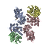
-Related structure data
| Related structure data | 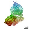 4598MC  6qnkC M: map data used to model this data C: citing same article ( |
|---|---|
| Similar structure data |


-Links
 PDBj
PDBj


- Assembly
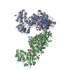
| Deposited unit | 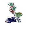
|
|---|---|
| 1 |
|
-Components
-Guanine nucleotide-binding protein ... , 3 types, 3 molecules ABG
| #1: Protein | Mass: 43182.078 Da / Num. of mol.: 1 Source method: isolated from a genetically manipulated source Source: (gene. exp.) Homo sapiens (human) / Gene: GNAI1 / Production host:  |
|---|---|
| #2: Protein | Mass: 37416.930 Da / Num. of mol.: 1 / Source method: isolated from a natural source / Source: (natural) |
| #3: Protein | Mass: 8556.918 Da / Num. of mol.: 1 / Source method: isolated from a natural source / Source: (natural) |
-Antibody , 2 types, 2 molecules LH
| #4: Antibody | Mass: 26309.551 Da / Num. of mol.: 1 Source method: isolated from a genetically manipulated source Source: (gene. exp.) |
|---|---|
| #5: Antibody | Mass: 26558.902 Da / Num. of mol.: 1 Source method: isolated from a genetically manipulated source Source: (gene. exp.) |
-Protein / Sugars / Non-polymers , 3 types, 3 molecules R
| #6: Protein | Mass: 39040.527 Da / Num. of mol.: 1 Source method: isolated from a genetically manipulated source Details: mutant N2C/M257Y/D282C / Source: (gene. exp.) Homo sapiens (human) / References: UniProt: P02699 |
|---|---|
| #7: Polysaccharide | 2-acetamido-2-deoxy-beta-D-glucopyranose-(1-4)-2-acetamido-2-deoxy-beta-D-glucopyranose Source method: isolated from a genetically manipulated source |
| #8: Chemical | ChemComp-RET /  Mass: 284.436 Da / Num. of mol.: 1 / Source method: obtained synthetically / Formula: C20H28O / Feature type: SUBJECT OF INVESTIGATION Mass: 284.436 Da / Num. of mol.: 1 / Source method: obtained synthetically / Formula: C20H28O / Feature type: SUBJECT OF INVESTIGATION |
-Details
| Has ligand of interest | Y |
|---|---|
| Has protein modification | Y |
-Experimental details
-Experiment
| Experiment | Method: ELECTRON MICROSCOPY |
|---|---|
| EM experiment | Aggregation state: PARTICLE / 3D reconstruction method: single particle reconstruction |
- Sample preparation
| Component |
| ||||||||||||||||||||||||||||||||||||
|---|---|---|---|---|---|---|---|---|---|---|---|---|---|---|---|---|---|---|---|---|---|---|---|---|---|---|---|---|---|---|---|---|---|---|---|---|---|
| Molecular weight | Value: 0.17 MDa / Experimental value: YES | ||||||||||||||||||||||||||||||||||||
| Source (natural) |
| ||||||||||||||||||||||||||||||||||||
| Source (recombinant) |
| ||||||||||||||||||||||||||||||||||||
| Buffer solution | pH: 7.5 Details: The detergent lauryl-maltose neopentyl glycol (LMNG) was used before the last purification step by gel filtration. In the gel filtration, detergent-free buffer was used. | ||||||||||||||||||||||||||||||||||||
| Buffer component |
| ||||||||||||||||||||||||||||||||||||
| Specimen | Conc.: 0.2 mg/ml / Embedding applied: NO / Shadowing applied: NO / Staining applied: NO / Vitrification applied: YES / Details: The sample was monodisperse. | ||||||||||||||||||||||||||||||||||||
| Specimen support | Grid material: COPPER / Grid mesh size: 300 divisions/in. | ||||||||||||||||||||||||||||||||||||
| Vitrification | Instrument: FEI VITROBOT MARK IV / Cryogen name: ETHANE / Humidity: 90 % / Chamber temperature: 295 K |
- Electron microscopy imaging
| Experimental equipment |  Model: Titan Krios / Image courtesy: FEI Company |
|---|---|
| Microscopy | Model: FEI TITAN KRIOS |
| Electron gun | Electron source:  FIELD EMISSION GUN / Accelerating voltage: 300 kV / Illumination mode: FLOOD BEAM FIELD EMISSION GUN / Accelerating voltage: 300 kV / Illumination mode: FLOOD BEAM |
| Electron lens | Mode: BRIGHT FIELD / Nominal magnification: 165000 X / Calibrated magnification: 165000 X / Nominal defocus max: 25000 nm / Nominal defocus min: 15000 nm / Calibrated defocus min: 15000 nm / Calibrated defocus max: 25000 nm / Cs: 2.7 mm / C2 aperture diameter: 100 µm / Alignment procedure: BASIC |
| Specimen holder | Cryogen: NITROGEN / Specimen holder model: FEI TITAN KRIOS AUTOGRID HOLDER / Temperature (max): 93 K / Temperature (min): 70 K |
| Image recording | Average exposure time: 10 sec. / Electron dose: 60 e/Å2 / Detector mode: SUPER-RESOLUTION / Film or detector model: GATAN K2 SUMMIT (4k x 4k) / Num. of grids imaged: 1 / Num. of real images: 3200 |
| EM imaging optics | Energyfilter name: GIF Quantum LS / Energyfilter slit width: 20 eV |
| Image scans | Sampling size: 5 µm / Width: 7676 / Height: 7420 / Movie frames/image: 40 / Used frames/image: 1-40 |
- Processing
| Software | Name: PHENIX / Version: 1.13_2998: / Classification: refinement | ||||||||||||||||||||||||||||||||||||||||
|---|---|---|---|---|---|---|---|---|---|---|---|---|---|---|---|---|---|---|---|---|---|---|---|---|---|---|---|---|---|---|---|---|---|---|---|---|---|---|---|---|---|
| EM software |
| ||||||||||||||||||||||||||||||||||||||||
| Image processing | Details: super resolution mode | ||||||||||||||||||||||||||||||||||||||||
| CTF correction | Type: NONE | ||||||||||||||||||||||||||||||||||||||||
| Particle selection | Num. of particles selected: 580000 | ||||||||||||||||||||||||||||||||||||||||
| Symmetry | Point symmetry: C1 (asymmetric) | ||||||||||||||||||||||||||||||||||||||||
| 3D reconstruction | Resolution: 4.38 Å / Resolution method: FSC 0.143 CUT-OFF / Num. of particles: 115000 / Algorithm: FOURIER SPACE / Num. of class averages: 3 / Symmetry type: POINT | ||||||||||||||||||||||||||||||||||||||||
| Atomic model building | Protocol: RIGID BODY FIT / Space: REAL | ||||||||||||||||||||||||||||||||||||||||
| Atomic model building |
|