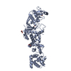
Evidence: gel filtration, Gel-filtrated sample shows the components for complex assembly in SDS-PAGE. It is a heterodimer of chains A and B in solution.
Type
Name
Symmetry operation
Number
identity operation
1_555
x,y,z
1
Buried area
2960 Å2
ΔGint
-10 kcal/mol
Surface area
22790 Å2
Method
PISA
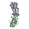
Unit cell
Length a, b, c (Å)
151.359, 151.359, 96.652
Angle α, β, γ (deg.)
90.000, 90.000, 120.000
Int Tables number
169
Space group name H-M
P61
-
Components
#1: Protein
Rhodopsin
Mass: 35800.941 Da / Num. of mol.: 1 Source method: isolated from a genetically manipulated source Source: (gene. exp.) Bos taurus (domestic cattle) / Gene: RHO / Cell line (production host): HEK293 / Production host: Homo sapiens (human) / References: UniProt: P02699
#2: Protein
Guaninenucleotide-bindingproteinG(o) subunitalpha
Mass: 24317.803 Da / Num. of mol.: 1 Source method: isolated from a genetically manipulated source Source: (gene. exp.) Homo sapiens (human) / Gene: GNAO1 / Production host: Escherichia coli (E. coli) / References: UniProt: P09471
In the structure databanks used in Yorodumi, some data are registered as the other names, "COVID-19 virus" and "2019-nCoV". Here are the details of the virus and the list of structure data.
Jan 31, 2019. EMDB accession codes are about to change! (news from PDBe EMDB page)
EMDB accession codes are about to change! (news from PDBe EMDB page)
The allocation of 4 digits for EMDB accession codes will soon come to an end. Whilst these codes will remain in use, new EMDB accession codes will include an additional digit and will expand incrementally as the available range of codes is exhausted. The current 4-digit format prefixed with “EMD-” (i.e. EMD-XXXX) will advance to a 5-digit format (i.e. EMD-XXXXX), and so on. It is currently estimated that the 4-digit codes will be depleted around Spring 2019, at which point the 5-digit format will come into force.
The EM Navigator/Yorodumi systems omit the EMD- prefix.
Related info.:Q: What is EMD? / ID/Accession-code notation in Yorodumi/EM Navigator
Yorodumi is a browser for structure data from EMDB, PDB, SASBDB, etc.
This page is also the successor to EM Navigator detail page, and also detail information page/front-end page for Omokage search.
The word "yorodu" (or yorozu) is an old Japanese word meaning "ten thousand". "mi" (miru) is to see.
Related info.:EMDB / PDB / SASBDB / Comparison of 3 databanks / Yorodumi Search / Aug 31, 2016. New EM Navigator & Yorodumi / Yorodumi Papers / Jmol/JSmol / Function and homology information / Changes in new EM Navigator and Yorodumi
 Movie
Movie Controller


 Open data
 Basic information
 Components
 Keywords
 Function and homology information
 Homo sapiens (human)
 X-RAY DIFFRACTION /
  Authors
 Switzerland,
  United Kingdom, 5items
  Citation
 Structure visualization
 Downloads & links
 Other downloads
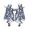


 PDBj
PDBj


 Assembly
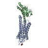


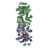
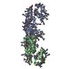
 Mass: 284.436 Da / Num. of mol.: 1 / Source method: obtained synthetically / Formula: C20H28O / Feature type: SUBJECT OF INVESTIGATION
 Type: D-saccharide, beta linking / Mass: 221.208 Da / Num. of mol.: 1
 Mass: 18.015 Da / Num. of mol.: 1 / Source method: isolated from a natural source / Formula: H2O
 Sample preparation
 Processing