| Entry | Database: PDB / ID: 5vyq
|
|---|
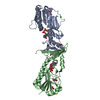
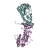
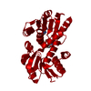
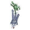
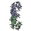
| Title | Crystal structure of the N-formyltransferase Rv3404c from mycobacterium tuberculosis in complex with YDP-Qui4N and folinic acid |
|---|
 Components Components | Uncharacterized protein |
|---|
 Keywords Keywords | TRANSFERASE / formyltransferase / deoxysugar |
|---|
| Function / homology |  Function and homology information Function and homology information
N-formyltransferase dimerization C-terminal domain / N-formyltransferase dimerization C-terminal domain / Formyl transferase, N-terminal domain / Formyl transferase, N-terminal / Formyl transferase / Formyl transferase, N-terminal domain superfamily / Rossmann fold / 3-Layer(aba) Sandwich / Alpha BetaSimilarity search - Domain/homology dTDP-4-amino-4,6-dideoxyglucose / Chem-EP1 / Chem-FON / : / : / THYMIDINE-5'-DIPHOSPHATE / dTDP-4-amino-4,6-dideoxyglucose formyltransferaseSimilarity search - Component |
|---|
| Biological species |  Mycobacterium tuberculosis CAS/NITR204 (bacteria) Mycobacterium tuberculosis CAS/NITR204 (bacteria) |
|---|
| Method |  X-RAY DIFFRACTION / MOLECULAR REPLACEMENT / Resolution: 1.6 Å X-RAY DIFFRACTION / MOLECULAR REPLACEMENT / Resolution: 1.6 Å |
|---|
 Authors Authors | Dunsirn, M.M. / Thoden, J.B. / Holden, H.M. |
|---|
| Funding support |  United States, 1items United States, 1items | Organization | Grant number | Country |
|---|
| National Institutes of Health/National Institute of General Medical Sciences (NIH/NIGMS) | GM115921 | United States |
|
|---|
 Citation Citation | Journal: Biochemistry / Year: 2017
Title: Biochemical Investigation of Rv3404c from Mycobacterium tuberculosis.
Authors: Dunsirn, M.M. / Thoden, J.B. / Gilbert, M. / Holden, H.M. |
|---|
| History | | Deposition | May 26, 2017 | Deposition site: RCSB / Processing site: RCSB |
|---|
| Revision 1.0 | Jul 12, 2017 | Provider: repository / Type: Initial release |
|---|
| Revision 1.1 | Aug 9, 2017 | Group: Database references / Category: citation
Item: _citation.journal_volume / _citation.page_first / _citation.page_last |
|---|
| Revision 1.2 | Sep 20, 2017 | Group: Author supporting evidence / Category: pdbx_audit_support / Item: _pdbx_audit_support.funding_organization |
|---|
| Revision 1.3 | Jan 1, 2020 | Group: Author supporting evidence / Category: pdbx_audit_support / Item: _pdbx_audit_support.funding_organization |
|---|
| Revision 1.4 | Oct 4, 2023 | Group: Data collection / Database references ...Data collection / Database references / Derived calculations / Refinement description
Category: chem_comp_atom / chem_comp_bond ...chem_comp_atom / chem_comp_bond / database_2 / pdbx_initial_refinement_model / pdbx_struct_conn_angle / struct_conn
Item: _database_2.pdbx_DOI / _database_2.pdbx_database_accession ..._database_2.pdbx_DOI / _database_2.pdbx_database_accession / _pdbx_struct_conn_angle.ptnr1_auth_asym_id / _pdbx_struct_conn_angle.ptnr1_auth_comp_id / _pdbx_struct_conn_angle.ptnr1_auth_seq_id / _pdbx_struct_conn_angle.ptnr1_label_asym_id / _pdbx_struct_conn_angle.ptnr1_label_atom_id / _pdbx_struct_conn_angle.ptnr1_label_comp_id / _pdbx_struct_conn_angle.ptnr1_label_seq_id / _pdbx_struct_conn_angle.ptnr1_symmetry / _pdbx_struct_conn_angle.ptnr2_auth_asym_id / _pdbx_struct_conn_angle.ptnr2_auth_comp_id / _pdbx_struct_conn_angle.ptnr2_auth_seq_id / _pdbx_struct_conn_angle.ptnr2_label_asym_id / _pdbx_struct_conn_angle.ptnr2_label_atom_id / _pdbx_struct_conn_angle.ptnr2_label_comp_id / _pdbx_struct_conn_angle.ptnr2_symmetry / _pdbx_struct_conn_angle.ptnr3_auth_asym_id / _pdbx_struct_conn_angle.ptnr3_auth_comp_id / _pdbx_struct_conn_angle.ptnr3_auth_seq_id / _pdbx_struct_conn_angle.ptnr3_label_asym_id / _pdbx_struct_conn_angle.ptnr3_label_atom_id / _pdbx_struct_conn_angle.ptnr3_label_comp_id / _pdbx_struct_conn_angle.ptnr3_label_seq_id / _pdbx_struct_conn_angle.ptnr3_symmetry / _pdbx_struct_conn_angle.value / _struct_conn.pdbx_dist_value / _struct_conn.ptnr1_auth_asym_id / _struct_conn.ptnr1_auth_comp_id / _struct_conn.ptnr1_auth_seq_id / _struct_conn.ptnr1_label_asym_id / _struct_conn.ptnr1_label_atom_id / _struct_conn.ptnr1_label_comp_id / _struct_conn.ptnr1_label_seq_id / _struct_conn.ptnr1_symmetry / _struct_conn.ptnr2_auth_asym_id / _struct_conn.ptnr2_auth_comp_id / _struct_conn.ptnr2_auth_seq_id / _struct_conn.ptnr2_label_asym_id / _struct_conn.ptnr2_label_atom_id / _struct_conn.ptnr2_label_comp_id / _struct_conn.ptnr2_label_seq_id / _struct_conn.ptnr2_symmetry |
|---|
|
|---|
 Movie
Movie Controller

 Yorodumi
 Open data
 Basic information
 Structure visualization
 Downloads & links
 Other downloads
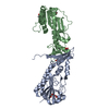


 PDBj
PDBj
 Assembly
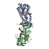

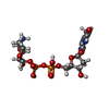


 Mass: 473.439 Da / Num. of mol.: 1 / Source method: obtained synthetically / Formula: C20H23N7O7
 Mass: 547.345 Da / Num. of mol.: 1 / Source method: obtained synthetically / Formula: C16H27N3O14P2
 Mass: 62.068 Da / Num. of mol.: 4 / Source method: obtained synthetically / Formula: C2H6O2
 Mass: 22.990 Da / Num. of mol.: 4 / Source method: obtained synthetically / Formula: Na
 Mass: 35.453 Da / Num. of mol.: 3 / Source method: obtained synthetically / Formula: Cl
 Mass: 252.331 Da / Num. of mol.: 2 / Source method: obtained synthetically / Formula: C9H20N2O4S / Comment: pH buffer*YM
 Mass: 6.941 Da / Num. of mol.: 1 / Source method: obtained synthetically / Formula: Li
 Mass: 402.188 Da / Num. of mol.: 1 / Source method: obtained synthetically / Formula: C10H16N2O11P2
 Mass: 39.098 Da / Num. of mol.: 1 / Source method: obtained synthetically / Formula: K
 Sample preparation
 Processing