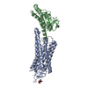
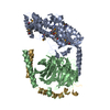
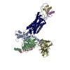
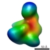
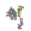
ジャーナル: Elife / 年: 2019 タイトル: Cryo-EM structure of the rhodopsin-Gαi-βγ complex reveals binding of the rhodopsin C-terminal tail to the gβ subunit. 著者: Ching-Ju Tsai / Jacopo Marino / Ricardo Adaixo / Filip Pamula / Jonas Muehle / Shoji Maeda / Tilman Flock / Nicholas Mi Taylor / Inayatulla Mohammed / Hugues Matile / Roger Jp Dawson / Xavier ...著者: Ching-Ju Tsai / Jacopo Marino / Ricardo Adaixo / Filip Pamula / Jonas Muehle / Shoji Maeda / Tilman Flock / Nicholas Mi Taylor / Inayatulla Mohammed / Hugues Matile / Roger Jp Dawson / Xavier Deupi / Henning Stahlberg / Gebhard Schertler / 要旨: One of the largest membrane protein families in eukaryotes are G protein-coupled receptors (GPCRs). GPCRs modulate cell physiology by activating diverse intracellular transducers, prominently ...One of the largest membrane protein families in eukaryotes are G protein-coupled receptors (GPCRs). GPCRs modulate cell physiology by activating diverse intracellular transducers, prominently heterotrimeric G proteins. The recent surge in structural data has expanded our understanding of GPCR-mediated signal transduction. However, many aspects, including the existence of transient interactions, remain elusive. We present the cryo-EM structure of the light-sensitive GPCR rhodopsin in complex with heterotrimeric Gi. Our density map reveals the receptor C-terminal tail bound to the Gβ subunit of the G protein, providing a structural foundation for the role of the C-terminal tail in GPCR signaling, and of Gβ as scaffold for recruiting Gα subunits and G protein-receptor kinases. By comparing available complexes, we found a small set of common anchoring points that are G protein-subtype specific. Taken together, our structure and analysis provide new structural basis for the molecular events of the GPCR signaling pathway.
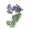
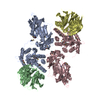
A: Guanine nucleotide-binding protein G(i) subunit alpha-1 B: Guanine nucleotide-binding protein G(I)/G(S)/G(T) subunit beta-1 G: Guanine nucleotide-binding protein G(T) subunit gamma-T1 L: Fab antibody fragment light chain H: Fab antibody fragment heavy chain R: Rhodopsin ヘテロ分子
根拠: gel filtration, The components of the complex were mixed., followed by gel filtration. The peak sample contains all the protein components, as evaluated by SDS-PAGE.
タイプ
名称
対称操作
数
identity operation
1_555
1
Buried area
14070 Å2
ΔGint
-70 kcal/mol
Surface area
61180 Å2
手法
PISA
-
要素
-
Guanine nucleotide-binding protein ... , 3種, 3分子 ABG
#1: タンパク質
Guaninenucleotide-bindingproteinG(i) subunitalpha-1 / Adenylate cyclase-inhibiting G alpha protein
分子量: 284.436 Da / 分子数: 1 / 由来タイプ: 合成 / 式: C20H28O / タイプ: SUBJECT OF INVESTIGATION
-
詳細
研究の焦点であるリガンドがあるか
Y
-
実験情報
-
実験
実験
手法: 電子顕微鏡法
EM実験
試料の集合状態: PARTICLE / 3次元再構成法: 単粒子再構成法
-
試料調製
構成要素
ID
名称
タイプ
Entity ID
Parent-ID
由来
1
Rhodopsin-Gi complex bound with antibody fragment Fab16
COMPLEX
#1-#6
0
MULTIPLESOURCES
2
Guanine nucleotide-binding protein alpha subunit
COMPLEX
#1
1
RECOMBINANT
3
Guanine nucleotide-binding protein beta/gamma subunit
COMPLEX
#2-#3
1
NATURAL
4
antibodyFABfragmentFab16
COMPLEX
#4-#5
1
RECOMBINANT
5
Rhodopsinロドプシン
COMPLEX
#6
1
RECOMBINANT
分子量
値: 0.17 MDa / 実験値: YES
由来(天然)
ID
Entity assembly-ID
生物種
Ncbi tax-ID
1
2
Homo sapiens (ヒト)
9606
2
3
Bos taurus (ウシ)
9913
3
4
Mus musculus (ハツカネズミ)
10090
4
5
Bos taurus (ウシ)
9913
由来(組換発現)
ID
Entity assembly-ID
生物種
Ncbi tax-ID
1
2
Escherichia coli BL21 (大腸菌)
511693
3
4
hybrid (その他)
37965
4
5
Homo sapiens (ヒト)
9606
緩衝液
pH: 7.5 詳細: The detergent lauryl-maltose neopentyl glycol (LMNG) was used before the last purification step by gel filtration. In the gel filtration, detergent-free buffer was used.
緩衝液成分
ID
濃度
名称
式
Buffer-ID
1
20mM
HEPES
C8H18N2O4S
1
2
150mM
Sodiumcholoride
NaCl塩化ナトリウム
1
試料
濃度: 0.2 mg/ml / 包埋: NO / シャドウイング: NO / 染色: NO / 凍結: YES / 詳細: The sample was monodisperse.
試料支持
グリッドの材料: COPPER / グリッドのサイズ: 300 divisions/in.
急速凍結
装置: FEI VITROBOT MARK IV / 凍結剤: ETHANE / 湿度: 90 % / 凍結前の試料温度: 295 K
 ムービー
ムービー コントローラー


 データを開く
 基本情報
基本情報 要素
要素 キーワード
キーワード SIGNALING PROTEIN / GPCR and G protein complex
 機能・相同性情報

 データ登録者
 スイス, 5件
  引用
引用 構造の表示
構造の表示 ダウンロードとリンク
 その他のダウンロード
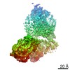


 PDBj
PDBj


 集合体
集合体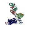


 分子量: 284.436 Da / 分子数: 1 / 由来タイプ: 合成 / 式: C20H28O / タイプ: SUBJECT OF INVESTIGATION
 試料調製
試料調製 電子顕微鏡撮影
 解析
解析