 Movie
Movie Controller


+ Open data

- Basic information
| Entry | Database: PDB / ID: 6qgq | ||||||
|---|---|---|---|---|---|---|---|
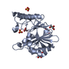
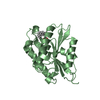
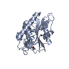
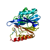
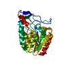
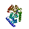
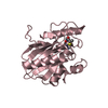
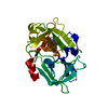
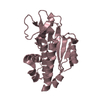
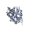
| Title | Crystal structure of APT1 C2S mutant bound to palmitic acid. | ||||||
 Components Components | (Acyl-protein thioesterase 1) x 2 | ||||||
 Keywords Keywords | HYDROLASE / acyl-protein thioesterase / palmitic acid / Depalmitoylation | ||||||
| Function / homology |  Function and homology information Function and homology informationprotein depalmitoylation / palmitoyl[protein] hydrolase / palmitoyl-(protein) hydrolase activity / negative regulation of aggrephagy / negative regulation of Golgi to plasma membrane protein transport / glycerophospholipase activity / lipase activity / Hydrolases; Acting on ester bonds; Thioester hydrolases / phosphatidylcholine lysophospholipase A1 activity / fatty acid transport ...protein depalmitoylation / palmitoyl[protein] hydrolase / palmitoyl-(protein) hydrolase activity / negative regulation of aggrephagy / negative regulation of Golgi to plasma membrane protein transport / glycerophospholipase activity / lipase activity / Hydrolases; Acting on ester bonds; Thioester hydrolases / phosphatidylcholine lysophospholipase A1 activity / fatty acid transport / eNOS activation / fatty acid metabolic process / RAS processing / nuclear membrane / endoplasmic reticulum / mitochondrion / extracellular exosome / nucleoplasm / plasma membrane / cytoplasm / cytosol Similarity search - Function | ||||||
| Biological species |  Homo sapiens (human) Homo sapiens (human) | ||||||
| Method |  X-RAY DIFFRACTION / SYNCHROTRON / MOLECULAR REPLACEMENT / Resolution: 2.601 Å X-RAY DIFFRACTION / SYNCHROTRON / MOLECULAR REPLACEMENT / Resolution: 2.601 Å | ||||||
 Authors Authors | Audagnotto, M. / Marcaida, M.J. / Ho, S. / Pojer, F. / Van der Goot, G. / Dal Peraro, M. | ||||||
| Funding support |  Switzerland, 1items Switzerland, 1items
| ||||||
 Citation Citation | Journal: Nat.Chem.Biol. / Year: 2021 Title: Palmitoylated acyl protein thioesterase APT2 deforms membranes to extract substrate acyl chains. Authors: Abrami, L. / Audagnotto, M. / Ho, S. / Marcaida, M.J. / Mesquita, F.S. / Anwar, M.U. / Sandoz, P.A. / Fonti, G. / Pojer, F. / Dal Peraro, M. / van der Goot, F.G. | ||||||
| History |
|
- Structure visualization
| Structure viewer | Molecule: MolmilJmol/JSmol |
|---|
- Downloads & links
-Download
| PDBx/mmCIF format | 6qgq.cif.gz | 180 KB | Display | PDBx/mmCIF format |
|---|---|---|---|---|
| PDB format | pdb6qgq.ent.gz | 143.2 KB | Display | PDB format |
| PDBx/mmJSON format | 6qgq.json.gz | Tree view | PDBx/mmJSON format | |
| Others |  Other downloads Other downloads |
-Validation report
| Arichive directory | https://data.pdbj.org/pub/pdb/validation_reports/qg/6qgqftp://data.pdbj.org/pub/pdb/validation_reports/qg/6qgq | HTTPS FTP |
|---|
-Related structure data
| Related structure data |  6qgnC  6qgoC  6qgsC  1fj2S C: citing same article ( S: Starting model for refinement |
|---|---|
| Similar structure data |
-Links
 PDBj
PDBj


- Assembly
| Deposited unit | 
| ||||||||
|---|---|---|---|---|---|---|---|---|---|
| 1 | 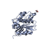
| ||||||||
| 2 | 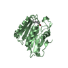
| ||||||||
| 3 | 
| ||||||||
| 4 | 
| ||||||||
| Unit cell |
|
-Components
| #1: Protein | Mass: 24977.805 Da / Num. of mol.: 3 / Mutation: C2S Source method: isolated from a genetically manipulated source Source: (gene. exp.) Homo sapiens (human) / Gene: LYPLA1, APT1, LPL1 / Production host:  References: UniProt: O75608, Hydrolases; Acting on ester bonds; Thioester hydrolases #2: Protein | | Mass: 25048.883 Da / Num. of mol.: 1 / Mutation: C2S Source method: isolated from a genetically manipulated source Source: (gene. exp.) Homo sapiens (human) / Gene: LYPLA1, APT1, LPL1 / Production host: References: UniProt: O75608, Hydrolases; Acting on ester bonds; Thioester hydrolases #3: Chemical | ChemComp-GOL /   Mass: 92.094 Da / Num. of mol.: 4 Mass: 92.094 Da / Num. of mol.: 4Source method: isolated from a genetically manipulated source Formula: C3H8O3 #4: Chemical |   Mass: 256.424 Da / Num. of mol.: 2 / Source method: isolated from a natural source / Formula: C16H32O2 Mass: 256.424 Da / Num. of mol.: 2 / Source method: isolated from a natural source / Formula: C16H32O2#5: Water | ChemComp-HOH / |  Mass: 18.015 Da / Num. of mol.: 132 / Source method: isolated from a natural source / Formula: H2O Mass: 18.015 Da / Num. of mol.: 132 / Source method: isolated from a natural source / Formula: H2O |
|---|
-Experimental details
-Experiment
| Experiment | Method: X-RAY DIFFRACTION / Number of used crystals: 1 |
|---|
- Sample preparation
| Crystal | Density Matthews: 2.51 Å3/Da / Density % sol: 50.9 % |
|---|---|
| Crystal grow | Temperature: 291 K / Method: vapor diffusion, hanging drop Details: 25% of of polyethylene glycol (PEG) 2000 MME, 0.3 M sodium acetate, 0.1 M sodium cacodylate pH 6.5; |
-Data collection
| Diffraction | Mean temperature: 100 K / Serial crystal experiment: N |
|---|---|
| Diffraction source | Source: SYNCHROTRON / Site: SLS / Beamline: X06DA / Wavelength: 1 Å |
| Detector | Type: DECTRIS PILATUS 2M-F / Detector: PIXEL / Date: Jun 27, 2016 |
| Radiation | Protocol: SINGLE WAVELENGTH / Monochromatic (M) / Laue (L): M / Scattering type: x-ray |
| Radiation wavelength | Wavelength: 1 Å / Relative weight: 1 |
| Reflection | Resolution: 2.6→48.71 Å / Num. obs: 30525 / % possible obs: 99.9 % / Redundancy: 7.9 % / Rmerge(I) obs: 0.18 / Net I/σ(I): 9.6 |
| Reflection shell | Resolution: 2.6→2.74 Å |
- Processing
| Software |
| ||||||||||||||||||||||||||||||||||||||||||||||||||||||||||||||||||||||||||||||||||||
|---|---|---|---|---|---|---|---|---|---|---|---|---|---|---|---|---|---|---|---|---|---|---|---|---|---|---|---|---|---|---|---|---|---|---|---|---|---|---|---|---|---|---|---|---|---|---|---|---|---|---|---|---|---|---|---|---|---|---|---|---|---|---|---|---|---|---|---|---|---|---|---|---|---|---|---|---|---|---|---|---|---|---|---|---|---|
| Refinement | Method to determine structure: MOLECULAR REPLACEMENT Starting model: 1FJ2 Resolution: 2.601→46.619 Å / SU ML: 0.26 / Cross valid method: FREE R-VALUE / σ(F): 1.34 / Phase error: 21.47
| ||||||||||||||||||||||||||||||||||||||||||||||||||||||||||||||||||||||||||||||||||||
| Solvent computation | Shrinkage radii: 0.9 Å / VDW probe radii: 1.11 Å | ||||||||||||||||||||||||||||||||||||||||||||||||||||||||||||||||||||||||||||||||||||
| Refinement step | Cycle: LAST / Resolution: 2.601→46.619 Å
| ||||||||||||||||||||||||||||||||||||||||||||||||||||||||||||||||||||||||||||||||||||
| Refine LS restraints |
| ||||||||||||||||||||||||||||||||||||||||||||||||||||||||||||||||||||||||||||||||||||
| LS refinement shell |
|
