- PDB-4f6z: Mutagenesis of zinc ligand residue Cys221 reveals plasticity in t... -
+
Open data
ID or keywords:
Loading...
-
Basic information
Entry
Database: PDB / ID: 4f6z
Title
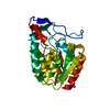
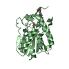
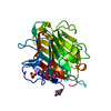
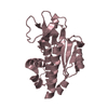
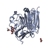
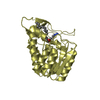
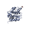
Mutagenesis of zinc ligand residue Cys221 reveals plasticity in the IMP-1 metallo-b-lactamase active site
Components
Beta-lactamase
Keywords
HYDROLASE / metallo-b-lactamase / b-lactamase
Function / homology
Function and homology information
antibiotic catabolic process / beta-lactamase activity / beta-lactamase / periplasmic space / response to antibiotic / zinc ion binding Similarity search - Function
Beta-lactamases class B signature 2. / Beta-lactamases class B signature 1. / Beta-lactamase, class-B, conserved site / : / : / Metallo-beta-lactamase superfamily / Ribonuclease Z/Hydroxyacylglutathione hydrolase-like / Metallo-beta-lactamase; Chain A / Metallo-beta-lactamase superfamily / Metallo-beta-lactamase ...Beta-lactamases class B signature 2. / Beta-lactamases class B signature 1. / Beta-lactamase, class-B, conserved site / : / : / Metallo-beta-lactamase superfamily / Ribonuclease Z/Hydroxyacylglutathione hydrolase-like / Metallo-beta-lactamase; Chain A / Metallo-beta-lactamase superfamily / Metallo-beta-lactamase / Ribonuclease Z/Hydroxyacylglutathione hydrolase-like / 4-Layer Sandwich / Alpha Beta Similarity search - Domain/homology
Mass: 18.015 Da / Num. of mol.: 151 / Source method: isolated from a natural source / Formula: H2O
-
Experimental details
-
Experiment
Experiment
Method: X-RAY DIFFRACTION / Number of used crystals: 1
-
Sample preparation
Crystal
Density Matthews: 2.39 Å3/Da / Density % sol: 48.52 %
Crystal grow
Temperature: 293 K / Method: vapor diffusion, hanging drop / pH: 7 Details: 25 mMTris-HCl containing 1 mM zinc sulfate at pH 7.0, and 25 mMTris-HCl with 1 mM zinc chloride at pH 7.0, and the mother liquor which consisted of 0.1 M tri-sodium citrate (pH 5.5) in 40% ...Details: 25 mMTris-HCl containing 1 mM zinc sulfate at pH 7.0, and 25 mMTris-HCl with 1 mM zinc chloride at pH 7.0, and the mother liquor which consisted of 0.1 M tri-sodium citrate (pH 5.5) in 40% PEG 600., VAPOR DIFFUSION, HANGING DROP, temperature 293K
In the structure databanks used in Yorodumi, some data are registered as the other names, "COVID-19 virus" and "2019-nCoV". Here are the details of the virus and the list of structure data.
Jan 31, 2019. EMDB accession codes are about to change! (news from PDBe EMDB page)
EMDB accession codes are about to change! (news from PDBe EMDB page)
The allocation of 4 digits for EMDB accession codes will soon come to an end. Whilst these codes will remain in use, new EMDB accession codes will include an additional digit and will expand incrementally as the available range of codes is exhausted. The current 4-digit format prefixed with “EMD-” (i.e. EMD-XXXX) will advance to a 5-digit format (i.e. EMD-XXXXX), and so on. It is currently estimated that the 4-digit codes will be depleted around Spring 2019, at which point the 5-digit format will come into force.
The EM Navigator/Yorodumi systems omit the EMD- prefix.
Related info.:Q: What is EMD? / ID/Accession-code notation in Yorodumi/EM Navigator
Yorodumi is a browser for structure data from EMDB, PDB, SASBDB, etc.
This page is also the successor to EM Navigator detail page, and also detail information page/front-end page for Omokage search.
The word "yorodu" (or yorozu) is an old Japanese word meaning "ten thousand". "mi" (miru) is to see.
Related info.:EMDB / PDB / SASBDB / Comparison of 3 databanks / Yorodumi Search / Aug 31, 2016. New EM Navigator & Yorodumi / Yorodumi Papers / Jmol/JSmol / Function and homology information / Changes in new EM Navigator and Yorodumi
 Movie
Movie Controller

 Yorodumi
 Open data
 Basic information
 Components
 Keywords
 Function and homology information
 Pseudomonas aeruginosa (bacteria)
 X-RAY DIFFRACTION /
  Authors
 Citation
 Structure visualization
 Downloads & links
 Other downloads
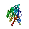


 PDBj
PDBj


 Assembly
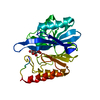

 Mass: 65.409 Da / Num. of mol.: 1 / Source method: obtained synthetically / Formula: Zn
 Mass: 189.100 Da / Num. of mol.: 1 / Source method: obtained synthetically / Formula: C6H5O7
 Mass: 18.015 Da / Num. of mol.: 151 / Source method: isolated from a natural source / Formula: H2O
 Sample preparation
 Processing