 Movie
Movie Controller

[English] 日本語
 Yorodumi
- PDB-6jyp: Crystal structure of uPA_H99Y in complex with 3-azanyl-5-(azepan-... -
+ Open data

- Basic information
| Entry | Database: PDB / ID: 6jyp | ||||||
|---|---|---|---|---|---|---|---|
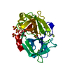
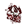
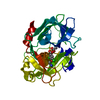
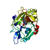
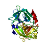
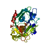
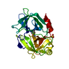
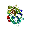
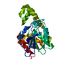
| Title | Crystal structure of uPA_H99Y in complex with 3-azanyl-5-(azepan-1-yl)-N-[bis(azanyl)methylidene]-6-chloranyl-pyrazine-2-carboxamide | ||||||
 Components Components | Urokinase-type plasminogen activator | ||||||
 Keywords Keywords | HYDROLASE / Urokinase / Amiloride | ||||||
| Function / homology |  Function and homology information Function and homology informationu-plasminogen activator / regulation of smooth muscle cell-matrix adhesion / urokinase plasminogen activator signaling pathway / regulation of plasminogen activation / regulation of integrin-mediated signaling pathway / regulation of fibrinolysis / protein complex involved in cell-matrix adhesion / regulation of wound healing / negative regulation of plasminogen activation / serine-type endopeptidase complex ...u-plasminogen activator / regulation of smooth muscle cell-matrix adhesion / urokinase plasminogen activator signaling pathway / regulation of plasminogen activation / regulation of integrin-mediated signaling pathway / regulation of fibrinolysis / protein complex involved in cell-matrix adhesion / regulation of wound healing / negative regulation of plasminogen activation / serine-type endopeptidase complex / regulation of smooth muscle cell migration / Dissolution of Fibrin Clot / regulation of cell adhesion mediated by integrin / smooth muscle cell migration / plasminogen activation / tertiary granule membrane / negative regulation of fibrinolysis / regulation of cell adhesion / serine protease inhibitor complex / specific granule membrane / fibrinolysis / positive regulation of epidermal growth factor receptor signaling pathway / chemotaxis / blood coagulation / regulation of cell population proliferation / response to hypoxia / positive regulation of cell migration / receptor ligand activity / serine-type endopeptidase activity / external side of plasma membrane / focal adhesion / Neutrophil degranulation / cell surface / signal transduction / proteolysis / : / extracellular exosome / extracellular region / plasma membrane Similarity search - Function | ||||||
| Biological species |  Homo sapiens (human) Homo sapiens (human) | ||||||
| Method |  X-RAY DIFFRACTION / SYNCHROTRON / MOLECULAR REPLACEMENT / Resolution: 2.25 Å X-RAY DIFFRACTION / SYNCHROTRON / MOLECULAR REPLACEMENT / Resolution: 2.25 Å | ||||||
 Authors Authors | Buckley, B. / Jiang, L.G. / Huang, M.D. / Kelso, M. / Ranson, M. | ||||||
| Funding support |  Australia, 1items Australia, 1items
| ||||||
 Citation Citation | Journal: To Be Published Title: uPA-HMA Authors: Buckley, B. / Jiang, L.G. / Huang, M.D. / Kelso, M. / Ranson, M. | ||||||
| History |
|
- Structure visualization
| Structure viewer | Molecule: MolmilJmol/JSmol |
|---|
- Downloads & links
-Download
| PDBx/mmCIF format | 6jyp.cif.gz | 65.9 KB | Display | PDBx/mmCIF format |
|---|---|---|---|---|
| PDB format | pdb6jyp.ent.gz | 46.6 KB | Display | PDB format |
| PDBx/mmJSON format | 6jyp.json.gz | Tree view | PDBx/mmJSON format | |
| Others |  Other downloads Other downloads |
-Validation report
| Arichive directory | https://data.pdbj.org/pub/pdb/validation_reports/jy/6jypftp://data.pdbj.org/pub/pdb/validation_reports/jy/6jyp | HTTPS FTP |
|---|
-Related structure data
| Related structure data |  6ag2C  6ag3C  6ag7C  6ag9C  4dvaS S: Starting model for refinement C: citing same article ( |
|---|---|
| Similar structure data |


-Links
 PDBj
PDBj

- Assembly
| Deposited unit | 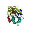
| ||||||||
|---|---|---|---|---|---|---|---|---|---|
| 1 |
| ||||||||
| Unit cell |
|
-Components
| #1: Protein | Mass: 27740.627 Da / Num. of mol.: 1 / Mutation: H99Y,C122A,N145Q Source method: isolated from a genetically manipulated source Source: (gene. exp.) Homo sapiens (human) / Gene: PLAU / Production host:  Komagataella pastoris (fungus) / References: UniProt: P00749, u-plasminogen activator Komagataella pastoris (fungus) / References: UniProt: P00749, u-plasminogen activator |
|---|---|
| #2: Chemical | ChemComp-HMX /   Mass: 311.771 Da / Num. of mol.: 1 / Source method: obtained synthetically / Formula: C12H18ClN7O / Feature type: SUBJECT OF INVESTIGATION Mass: 311.771 Da / Num. of mol.: 1 / Source method: obtained synthetically / Formula: C12H18ClN7O / Feature type: SUBJECT OF INVESTIGATION |
| #3: Chemical | ChemComp-SO4 /   Mass: 96.063 Da / Num. of mol.: 1 / Source method: obtained synthetically / Formula: SO4 Mass: 96.063 Da / Num. of mol.: 1 / Source method: obtained synthetically / Formula: SO4 |
| #4: Water | ChemComp-HOH /  Mass: 18.015 Da / Num. of mol.: 50 / Source method: isolated from a natural source / Formula: H2O Mass: 18.015 Da / Num. of mol.: 50 / Source method: isolated from a natural source / Formula: H2O |
| Has ligand of interest | Y |
| Has protein modification | Y |
-Experimental details
-Experiment
| Experiment | Method: X-RAY DIFFRACTION / Number of used crystals: 1 |
|---|
- Sample preparation
| Crystal | Density Matthews: 2.15 Å3/Da / Density % sol: 42.8 % |
|---|---|
| Crystal grow | Temperature: 298 K / Method: vapor diffusion, sitting drop Details: 0.05M sodium citrate at pH 4.6, 1.95M (NH4)2SO4, 0.03% NaN3, 5% PEG 400 |
-Data collection
| Diffraction | Mean temperature: 100 K / Serial crystal experiment: N |
|---|---|
| Diffraction source | Source: SYNCHROTRON / Site: NFPSS  / Beamline: BL19U1 / Wavelength: 1 Å / Beamline: BL19U1 / Wavelength: 1 Å |
| Detector | Type: DECTRIS PILATUS 6M / Detector: PIXEL / Date: Dec 13, 2013 |
| Radiation | Protocol: SINGLE WAVELENGTH / Monochromatic (M) / Laue (L): M / Scattering type: x-ray |
| Radiation wavelength | Wavelength: 1 Å / Relative weight: 1 |
| Reflection | Resolution: 2.25→60 Å / Num. obs: 10984 / % possible obs: 99.7 % / Redundancy: 3.5 % / Rmerge(I) obs: 0.104 / Net I/σ(I): 30 |
| Reflection shell | Resolution: 2.25→2.29 Å / Rmerge(I) obs: 0.691 / Num. unique obs: 559 |
- Processing
| Software |
| |||||||||||||||||||||||||||||||||||||||||||||||||||||||||||||||||||||||||||||||||||||||||||||||||||||||||||||||||||||||||||||||||||||||||||||||||
|---|---|---|---|---|---|---|---|---|---|---|---|---|---|---|---|---|---|---|---|---|---|---|---|---|---|---|---|---|---|---|---|---|---|---|---|---|---|---|---|---|---|---|---|---|---|---|---|---|---|---|---|---|---|---|---|---|---|---|---|---|---|---|---|---|---|---|---|---|---|---|---|---|---|---|---|---|---|---|---|---|---|---|---|---|---|---|---|---|---|---|---|---|---|---|---|---|---|---|---|---|---|---|---|---|---|---|---|---|---|---|---|---|---|---|---|---|---|---|---|---|---|---|---|---|---|---|---|---|---|---|---|---|---|---|---|---|---|---|---|---|---|---|---|---|---|---|
| Refinement | Method to determine structure: MOLECULAR REPLACEMENT Starting model: 4DVA Resolution: 2.25→30.33 Å / Cor.coef. Fo:Fc: 0.959 / Cor.coef. Fo:Fc free: 0.933 / SU B: 8.512 / SU ML: 0.21 / Cross valid method: THROUGHOUT / ESU R: 0.409 / ESU R Free: 0.255 / Details: HYDROGENS HAVE BEEN ADDED IN THE RIDING POSITIONS
| |||||||||||||||||||||||||||||||||||||||||||||||||||||||||||||||||||||||||||||||||||||||||||||||||||||||||||||||||||||||||||||||||||||||||||||||||
| Solvent computation | Ion probe radii: 0.8 Å / Shrinkage radii: 0.8 Å / VDW probe radii: 1.2 Å | |||||||||||||||||||||||||||||||||||||||||||||||||||||||||||||||||||||||||||||||||||||||||||||||||||||||||||||||||||||||||||||||||||||||||||||||||
| Displacement parameters | Biso max: 124.16 Å2 / Biso mean: 50.97 Å2 / Biso min: 26.42 Å2
| |||||||||||||||||||||||||||||||||||||||||||||||||||||||||||||||||||||||||||||||||||||||||||||||||||||||||||||||||||||||||||||||||||||||||||||||||
| Refinement step | Cycle: final / Resolution: 2.25→30.33 Å
| |||||||||||||||||||||||||||||||||||||||||||||||||||||||||||||||||||||||||||||||||||||||||||||||||||||||||||||||||||||||||||||||||||||||||||||||||
| Refine LS restraints |
| |||||||||||||||||||||||||||||||||||||||||||||||||||||||||||||||||||||||||||||||||||||||||||||||||||||||||||||||||||||||||||||||||||||||||||||||||
| LS refinement shell | Resolution: 2.25→2.3 Å / Rfactor Rfree error: 0 / Total num. of bins used: 20
|
