 Movie
Movie Controller

[English] 日本語
 Yorodumi
- PDB-6ooi: Crystal structure of triosephosphate isomerase from Schistosoma m... -
+ Open data

- Basic information
| Entry | Database: PDB / ID: 6ooi | ||||||
|---|---|---|---|---|---|---|---|
| Title | Crystal structure of triosephosphate isomerase from Schistosoma mansoni in complex with 2PG | ||||||
 Components Components | Triosephosphate isomerase | ||||||
 Keywords Keywords | ISOMERASE | ||||||
| Function / homology |  Function and homology information Function and homology informationtriose-phosphate isomerase / triose-phosphate isomerase activity / glyceraldehyde-3-phosphate biosynthetic process / glycerol catabolic process / glycolytic process / gluconeogenesis / cytosol Similarity search - Function | ||||||
| Biological species |  | ||||||
| Method |  X-RAY DIFFRACTION / MOLECULAR REPLACEMENT / Resolution: 2.14 Å X-RAY DIFFRACTION / MOLECULAR REPLACEMENT / Resolution: 2.14 Å | ||||||
 Authors Authors | Jimenez-Sandoval, P. / Brieba, L. | ||||||
| Funding support |  Mexico, 1items Mexico, 1items
| ||||||
 Citation Citation | Journal: Plos Negl Trop Dis / Year: 2020 Title: Crystal structures of Triosephosphate Isomerases from Taenia solium and Schistosoma mansoni provide insights for vaccine rationale and drug design against helminth parasites. Authors: Jimenez-Sandoval, P. / Castro-Torres, E. / Gonzalez-Gonzalez, R. / Diaz-Quezada, C. / Gurrola, M. / Camacho-Manriquez, L.D. / Leyva-Navarro, L. / Brieba, L.G. | ||||||
| History |
|
- Structure visualization
| Structure viewer | Molecule: MolmilJmol/JSmol |
|---|
- Downloads & links
-Download
| PDBx/mmCIF format | 6ooi.cif.gz | 407.5 KB | Display | PDBx/mmCIF format |
|---|---|---|---|---|
| PDB format | pdb6ooi.ent.gz | 327.8 KB | Display | PDB format |
| PDBx/mmJSON format | 6ooi.json.gz | Tree view | PDBx/mmJSON format | |
| Others |  Other downloads Other downloads |
-Validation report
| Arichive directory | https://data.pdbj.org/pub/pdb/validation_reports/oo/6ooiftp://data.pdbj.org/pub/pdb/validation_reports/oo/6ooi | HTTPS FTP |
|---|
-Related structure data
| Related structure data |  6oogC  5zfxS S: Starting model for refinement C: citing same article ( |
|---|---|
| Similar structure data |


-Links
 PDBj
PDBj
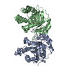
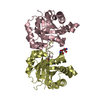
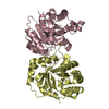
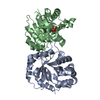
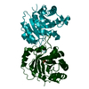
- Assembly
| Deposited unit | 
| ||||||||||
|---|---|---|---|---|---|---|---|---|---|---|---|
| 1 | 
| ||||||||||
| 2 | 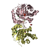
| ||||||||||
| 3 | 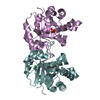
| ||||||||||
| 4 | 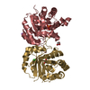
| ||||||||||
| Unit cell |
|
-Components
-Protein , 1 types, 8 molecules ABCDEFGH
| #1: Protein | Mass: 28456.465 Da / Num. of mol.: 8 Source method: isolated from a genetically manipulated source Source: (gene. exp.)  |
|---|
-Non-polymers , 5 types, 1099 molecules 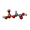


| #2: Chemical | ChemComp-PGA /  Mass: 156.031 Da / Num. of mol.: 8 / Source method: obtained synthetically / Formula: C2H5O6P Mass: 156.031 Da / Num. of mol.: 8 / Source method: obtained synthetically / Formula: C2H5O6P#3: Chemical | ChemComp-CL /  Mass: 35.453 Da / Num. of mol.: 6 / Source method: isolated from a natural source / Formula: Cl Mass: 35.453 Da / Num. of mol.: 6 / Source method: isolated from a natural source / Formula: Cl#4: Chemical | ChemComp-NA /  Mass: 22.990 Da / Num. of mol.: 8 / Source method: obtained synthetically / Formula: Na Mass: 22.990 Da / Num. of mol.: 8 / Source method: obtained synthetically / Formula: Na#5: Chemical |  Mass: 92.094 Da / Num. of mol.: 2 Mass: 92.094 Da / Num. of mol.: 2Source method: isolated from a genetically manipulated source Formula: C3H8O3 #6: Water | ChemComp-HOH / | Mass: 18.015 Da / Num. of mol.: 1075 / Source method: isolated from a natural source / Formula: H2O |
|---|
-Details
| Has ligand of interest | N |
|---|
-Experimental details
-Experiment
| Experiment | Method: X-RAY DIFFRACTION / Number of used crystals: 1 |
|---|
- Sample preparation
| Crystal | Density Matthews: 2.4 Å3/Da / Density % sol: 48.79 % |
|---|---|
| Crystal grow | Temperature: 292 K / Method: vapor diffusion, hanging drop / pH: 6.5 Details: 0.05M Cesium chloride 0.1M MES monohydrate 6.5 30% v/v Jeffamine M-600 |
-Data collection
| Diffraction | Mean temperature: 100 K / Serial crystal experiment: N |
|---|---|
| Diffraction source | Source: ROTATING ANODE / Type: RIGAKU MICROMAX-002+ / Wavelength: 1.5419 Å |
| Detector | Type: RIGAKU RAXIS IV++ / Detector: IMAGE PLATE / Date: Aug 8, 2018 |
| Radiation | Protocol: SINGLE WAVELENGTH / Monochromatic (M) / Laue (L): M / Scattering type: x-ray |
| Radiation wavelength | Wavelength: 1.5419 Å / Relative weight: 1 |
| Reflection | Resolution: 2.14→58.61 Å / Num. obs: 115018 / % possible obs: 100 % / Redundancy: 4.5 % / CC1/2: 0.984 / Rmerge(I) obs: 0.153 / Rpim(I) all: 0.081 / Rrim(I) all: 0.173 / Net I/σ(I): 5.6 / Num. measured all: 517652 / Scaling rejects: 76 |
| Reflection shell | Resolution: 2.14→2.18 Å / Redundancy: 4.2 % / Rmerge(I) obs: 0.804 / Num. measured all: 24134 / Num. unique obs: 5688 / CC1/2: 0.639 / Rpim(I) all: 0.436 / Rrim(I) all: 0.917 / Net I/σ(I) obs: 1.6 / % possible all: 100 |
- Processing
| Software |
| |||||||||||||||||||||||||||||||||||||||||||||||||||||||||||||||||||||||||||||||||||||||||||||||||||||||||
|---|---|---|---|---|---|---|---|---|---|---|---|---|---|---|---|---|---|---|---|---|---|---|---|---|---|---|---|---|---|---|---|---|---|---|---|---|---|---|---|---|---|---|---|---|---|---|---|---|---|---|---|---|---|---|---|---|---|---|---|---|---|---|---|---|---|---|---|---|---|---|---|---|---|---|---|---|---|---|---|---|---|---|---|---|---|---|---|---|---|---|---|---|---|---|---|---|---|---|---|---|---|---|---|---|---|---|
| Refinement | Method to determine structure: MOLECULAR REPLACEMENT Starting model: 5ZFX Resolution: 2.14→58.609 Å / SU ML: 0.24 / Cross valid method: THROUGHOUT / σ(F): 1.35 / Phase error: 22.62 / Stereochemistry target values: ML
| |||||||||||||||||||||||||||||||||||||||||||||||||||||||||||||||||||||||||||||||||||||||||||||||||||||||||
| Solvent computation | Shrinkage radii: 0.9 Å / VDW probe radii: 1.11 Å / Solvent model: FLAT BULK SOLVENT MODEL | |||||||||||||||||||||||||||||||||||||||||||||||||||||||||||||||||||||||||||||||||||||||||||||||||||||||||
| Displacement parameters | Biso max: 69.04 Å2 / Biso mean: 24.1252 Å2 / Biso min: 7.17 Å2 | |||||||||||||||||||||||||||||||||||||||||||||||||||||||||||||||||||||||||||||||||||||||||||||||||||||||||
| Refinement step | Cycle: final / Resolution: 2.14→58.609 Å
| |||||||||||||||||||||||||||||||||||||||||||||||||||||||||||||||||||||||||||||||||||||||||||||||||||||||||
| LS refinement shell | Refine-ID: X-RAY DIFFRACTION / Rfactor Rfree error: 0 / Total num. of bins used: 14
|
