 Movie
Movie Controller

[English] 日本語
 Yorodumi
- PDB-5zfx: Crystal Structure of Triosephosphate isomerase from Opisthorchis ... -
+ Open data

- Basic information
| Entry | Database: PDB / ID: 5zfx | ||||||
|---|---|---|---|---|---|---|---|
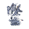
| Title | Crystal Structure of Triosephosphate isomerase from Opisthorchis viverrini | ||||||
 Components Components | Triosephosphate isomerase | ||||||
 Keywords Keywords | ISOMERASE / Tim-barrel / Triosephosphate isomerase | ||||||
| Function / homology |  Function and homology information Function and homology informationtriose-phosphate isomerase / triose-phosphate isomerase activity / glyceraldehyde-3-phosphate biosynthetic process / glycerol catabolic process / gluconeogenesis / glycolytic process / metal ion binding / cytosol Similarity search - Function | ||||||
| Biological species |  Opisthorchis viverrini (Southeast Asian liver fluke) Opisthorchis viverrini (Southeast Asian liver fluke) | ||||||
| Method |  X-RAY DIFFRACTION / SYNCHROTRON / MOLECULAR REPLACEMENT / Resolution: 1.751 Å X-RAY DIFFRACTION / SYNCHROTRON / MOLECULAR REPLACEMENT / Resolution: 1.751 Å | ||||||
 Authors Authors | Son, J. / Kim, S. / Kim, S.E. / Lee, H. / Lee, M.R. / Hwang, K.Y. | ||||||
 Citation Citation | Journal: Sci Rep / Year: 2018 Title: Structural Analysis of an Epitope Candidate of Triosephosphate Isomerase in Opisthorchis viverrini. Authors: Son, J. / Kim, S. / Kim, S.E. / Lee, H. / Lee, M.R. / Hwang, K.Y. | ||||||
| History |
|
- Structure visualization
| Structure viewer | Molecule: MolmilJmol/JSmol |
|---|
- Downloads & links
-Download
| PDBx/mmCIF format | 5zfx.cif.gz | 228.8 KB | Display | PDBx/mmCIF format |
|---|---|---|---|---|
| PDB format | pdb5zfx.ent.gz | 179.4 KB | Display | PDB format |
| PDBx/mmJSON format | 5zfx.json.gz | Tree view | PDBx/mmJSON format | |
| Others |  Other downloads Other downloads |
-Validation report
| Arichive directory | https://data.pdbj.org/pub/pdb/validation_reports/zf/5zfxftp://data.pdbj.org/pub/pdb/validation_reports/zf/5zfx | HTTPS FTP |
|---|
-Related structure data
| Related structure data | 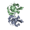 5zg4C 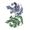 5zg5C 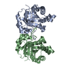 5zgaC  1tphS S: Starting model for refinement C: citing same article ( |
|---|---|
| Similar structure data |


-Links
 PDBj
PDBj
- Assembly
| Deposited unit | 
| ||||||||||||||||||||||||||||||||||||||||||||||||||||||||||||||||||||||||||||||
|---|---|---|---|---|---|---|---|---|---|---|---|---|---|---|---|---|---|---|---|---|---|---|---|---|---|---|---|---|---|---|---|---|---|---|---|---|---|---|---|---|---|---|---|---|---|---|---|---|---|---|---|---|---|---|---|---|---|---|---|---|---|---|---|---|---|---|---|---|---|---|---|---|---|---|---|---|---|---|---|
| 1 | 
| ||||||||||||||||||||||||||||||||||||||||||||||||||||||||||||||||||||||||||||||
| 2 | 
| ||||||||||||||||||||||||||||||||||||||||||||||||||||||||||||||||||||||||||||||
| Unit cell |
| ||||||||||||||||||||||||||||||||||||||||||||||||||||||||||||||||||||||||||||||
| Noncrystallographic symmetry (NCS) | NCS domain:
NCS domain segments: Ens-ID: 1 / End auth comp-ID: ALA / End label comp-ID: ALA
|
-Components
| #1: Protein | Mass: 30008.338 Da / Num. of mol.: 4 Source method: isolated from a genetically manipulated source Source: (gene. exp.) Opisthorchis viverrini (Southeast Asian liver fluke)Gene: T265_10017 / Plasmid: pET17b / Production host:  #2: Chemical |   Mass: 24.305 Da / Num. of mol.: 2 / Source method: obtained synthetically / Formula: Mg Mass: 24.305 Da / Num. of mol.: 2 / Source method: obtained synthetically / Formula: Mg#3: Water | ChemComp-HOH / |  Mass: 18.015 Da / Num. of mol.: 1195 / Source method: isolated from a natural source / Formula: H2O Mass: 18.015 Da / Num. of mol.: 1195 / Source method: isolated from a natural source / Formula: H2OSequence details | The sequence conflicts may be resulted from the difference of subspecies of the source organism. | |
|---|
-Experimental details
-Experiment
| Experiment | Method: X-RAY DIFFRACTION / Number of used crystals: 1 |
|---|
- Sample preparation
| Crystal | Density Matthews: 2.22 Å3/Da / Density % sol: 39.6 % / Mosaicity: 0.508 ° |
|---|---|
| Crystal grow | Temperature: 293 K / Method: evaporation / pH: 8.5 / Details: PEG 3350 |
-Data collection
| Diffraction | Mean temperature: 273 K | |||||||||||||||||||||||||||||||||||||||||||||||||||||||||||||||||||||||||||||
|---|---|---|---|---|---|---|---|---|---|---|---|---|---|---|---|---|---|---|---|---|---|---|---|---|---|---|---|---|---|---|---|---|---|---|---|---|---|---|---|---|---|---|---|---|---|---|---|---|---|---|---|---|---|---|---|---|---|---|---|---|---|---|---|---|---|---|---|---|---|---|---|---|---|---|---|---|---|---|
| Diffraction source | Source: SYNCHROTRON / Site: PAL/PLS  / Beamline: 5C (4A) / Wavelength: 1.00001 Å / Beamline: 5C (4A) / Wavelength: 1.00001 Å | |||||||||||||||||||||||||||||||||||||||||||||||||||||||||||||||||||||||||||||
| Detector | Type: ADSC QUANTUM 315r / Detector: CCD / Date: Nov 6, 2013 | |||||||||||||||||||||||||||||||||||||||||||||||||||||||||||||||||||||||||||||
| Radiation | Protocol: SINGLE WAVELENGTH / Monochromatic (M) / Laue (L): M / Scattering type: x-ray | |||||||||||||||||||||||||||||||||||||||||||||||||||||||||||||||||||||||||||||
| Radiation wavelength | Wavelength: 1.00001 Å / Relative weight: 1 | |||||||||||||||||||||||||||||||||||||||||||||||||||||||||||||||||||||||||||||
| Reflection | Resolution: 1.75→50 Å / Num. obs: 96825 / % possible obs: 98.8 % / Redundancy: 5.6 % / Biso Wilson estimate: 15.65 Å2 / Rmerge(I) obs: 0.105 / Χ2: 11.73 / Net I/σ(I): 37.7 / Num. measured all: 541897 | |||||||||||||||||||||||||||||||||||||||||||||||||||||||||||||||||||||||||||||
| Reflection shell |
|
- Processing
| Software |
| |||||||||||||||||||||||||||||||||||||||||||||||||||||||||||||||||||||||||||||||||||||||||||||||||||||||||
|---|---|---|---|---|---|---|---|---|---|---|---|---|---|---|---|---|---|---|---|---|---|---|---|---|---|---|---|---|---|---|---|---|---|---|---|---|---|---|---|---|---|---|---|---|---|---|---|---|---|---|---|---|---|---|---|---|---|---|---|---|---|---|---|---|---|---|---|---|---|---|---|---|---|---|---|---|---|---|---|---|---|---|---|---|---|---|---|---|---|---|---|---|---|---|---|---|---|---|---|---|---|---|---|---|---|---|
| Refinement | Method to determine structure: MOLECULAR REPLACEMENT Starting model: 1TPH Resolution: 1.751→44.075 Å / SU ML: 0.16 / Cross valid method: THROUGHOUT / σ(F): 1.56 / Phase error: 18.42 / Stereochemistry target values: ML
| |||||||||||||||||||||||||||||||||||||||||||||||||||||||||||||||||||||||||||||||||||||||||||||||||||||||||
| Solvent computation | Shrinkage radii: 0.9 Å / VDW probe radii: 1.11 Å / Solvent model: FLAT BULK SOLVENT MODEL | |||||||||||||||||||||||||||||||||||||||||||||||||||||||||||||||||||||||||||||||||||||||||||||||||||||||||
| Displacement parameters | Biso max: 87.76 Å2 / Biso mean: 21.1921 Å2 / Biso min: 5.06 Å2 | |||||||||||||||||||||||||||||||||||||||||||||||||||||||||||||||||||||||||||||||||||||||||||||||||||||||||
| Refinement step | Cycle: final / Resolution: 1.751→44.075 Å
| |||||||||||||||||||||||||||||||||||||||||||||||||||||||||||||||||||||||||||||||||||||||||||||||||||||||||
| Refine LS restraints |
| |||||||||||||||||||||||||||||||||||||||||||||||||||||||||||||||||||||||||||||||||||||||||||||||||||||||||
| Refine LS restraints NCS |
| |||||||||||||||||||||||||||||||||||||||||||||||||||||||||||||||||||||||||||||||||||||||||||||||||||||||||
| LS refinement shell | Refine-ID: X-RAY DIFFRACTION / Rfactor Rfree error: 0 / Total num. of bins used: 14
|
