 Movie
Movie Controller

[English] 日本語
 Yorodumi
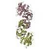
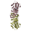
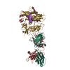
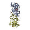
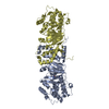
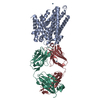
- PDB-6ofi: CRYSTAL STRUCTURE OF the RV144 C1-C2 SPECIFIC ANTIBODY CH55 FAB I... -
+ Open data

- Basic information
| Entry | Database: PDB / ID: 6ofi | ||||||||||||
|---|---|---|---|---|---|---|---|---|---|---|---|---|---|
| Title | CRYSTAL STRUCTURE OF the RV144 C1-C2 SPECIFIC ANTIBODY CH55 FAB IN COMPLEX WITH HIV-1 CLADE A/E GP120 | ||||||||||||
 Components Components |
| ||||||||||||
 Keywords Keywords | IMMUNE SYSTEM / ANTI-HIV-1 ENV ANTIBODY CH55 / CD4I ANTIBODY / ADCC / HIV-1 ENV / RV144 VACCINE TRIAL / CLADE A/E 93TH057 / VIRAL PROTEIN-IMMUNE SYSTEM COMPLEX | ||||||||||||
| Function / homology |  Function and homology information Function and homology informationmembrane fusion involved in viral entry into host cell / viral envelope / symbiont entry into host cell / virion attachment to host cell / virion membrane Similarity search - Function | ||||||||||||
| Biological species |   Human immunodeficiency virus 1 Human immunodeficiency virus 1 Homo sapiens (human) Homo sapiens (human) | ||||||||||||
| Method |  X-RAY DIFFRACTION / SYNCHROTRON / MOLECULAR REPLACEMENT / Resolution: 3.85 Å X-RAY DIFFRACTION / SYNCHROTRON / MOLECULAR REPLACEMENT / Resolution: 3.85 Å | ||||||||||||
 Authors Authors | Tolbert, W.D. / Yan, F. / Van, V. / Pazgier, M. | ||||||||||||
| Funding support |  United States, 3items United States, 3items
| ||||||||||||
 Citation Citation | Journal: Mbio / Year: 2020 Title: Recognition Patterns of the C1/C2 Epitopes Involved in Fc-Mediated Response in HIV-1 Natural Infection and the RV114 Vaccine Trial. Authors: Tolbert, W.D. / Van, V. / Sherburn, R. / Tuyishime, M. / Yan, F. / Nguyen, D.N. / Stanfield-Oakley, S. / Easterhoff, D. / Bonsignori, M. / Haynes, B.F. / Moody, M.A. / Ray, K. / Ferrari, G. ...Authors: Tolbert, W.D. / Van, V. / Sherburn, R. / Tuyishime, M. / Yan, F. / Nguyen, D.N. / Stanfield-Oakley, S. / Easterhoff, D. / Bonsignori, M. / Haynes, B.F. / Moody, M.A. / Ray, K. / Ferrari, G. / Lewis, G.K. / Pazgier, M. | ||||||||||||
| History |
|
- Structure visualization
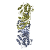
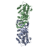
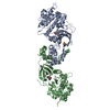
| Structure viewer | Molecule: MolmilJmol/JSmol |
|---|
- Downloads & links
-Download
| PDBx/mmCIF format | 6ofi.cif.gz | 309.3 KB | Display | PDBx/mmCIF format |
|---|---|---|---|---|
| PDB format | pdb6ofi.ent.gz | 252 KB | Display | PDB format |
| PDBx/mmJSON format | 6ofi.json.gz | Tree view | PDBx/mmJSON format | |
| Others |  Other downloads Other downloads |
-Validation report
| Arichive directory | https://data.pdbj.org/pub/pdb/validation_reports/of/6ofiftp://data.pdbj.org/pub/pdb/validation_reports/of/6ofi | HTTPS FTP |
|---|
-Related structure data
| Related structure data | 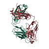 4fz8C 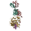 6mg7C 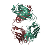 6oedSC 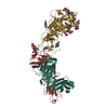 6oejC  3tgtS S: Starting model for refinement C: citing same article ( |
|---|---|
| Similar structure data |


-Links
 PDBj
PDBj


- Assembly
| Deposited unit | 
| ||||||||
|---|---|---|---|---|---|---|---|---|---|
| 1 |
| ||||||||
| Unit cell |
|
-Components
| #1: Protein | Mass: 39356.613 Da / Num. of mol.: 1 / Mutation: H375S Source method: isolated from a genetically manipulated source Source: (gene. exp.) Human immunodeficiency virus 1 / Gene: HIV-1 Env / Cell (production host): HEK 293 GnT1- / Production host: Homo sapiens (human) / References: UniProt: A0A0M3KKW9 | ||
|---|---|---|---|
| #2: Antibody | Mass: 24004.059 Da / Num. of mol.: 1 Source method: isolated from a genetically manipulated source Source: (gene. exp.) Homo sapiens (human) / Cell (production host): HEK 293 / Production host: Homo sapiens (human) | ||
| #3: Antibody | Mass: 23444.938 Da / Num. of mol.: 1 Source method: isolated from a genetically manipulated source Source: (gene. exp.) Homo sapiens (human) / Cell (production host): HEK 293 / Production host: Homo sapiens (human) | ||
| #4: Sugar | ChemComp-NAG /   Type: D-saccharide, beta linking / Mass: 221.208 Da / Num. of mol.: 10 Type: D-saccharide, beta linking / Mass: 221.208 Da / Num. of mol.: 10Source method: isolated from a genetically manipulated source Formula: C8H15NO6 Has protein modification | Y | |
-Experimental details
-Experiment
| Experiment | Method: X-RAY DIFFRACTION / Number of used crystals: 1 |
|---|
- Sample preparation
| Crystal | Density Matthews: 2.79 Å3/Da / Density % sol: 55.97 % |
|---|---|
| Crystal grow | Temperature: 294 K / Method: vapor diffusion, hanging drop / pH: 5.5 / Details: 15% PEG 6000, 0.1 M sodium citrate pH 5.5 |
-Data collection
| Diffraction | Mean temperature: 100 K / Serial crystal experiment: N |
|---|---|
| Diffraction source | Source: SYNCHROTRON / Site: SSRL / Beamline: BL12-2 / Wavelength: 0.97946 Å |
| Detector | Type: DECTRIS PILATUS 6M / Detector: PIXEL / Date: May 28, 2018 |
| Radiation | Monochromator: Si 111 / Protocol: SINGLE WAVELENGTH / Monochromatic (M) / Laue (L): M / Scattering type: x-ray |
| Radiation wavelength | Wavelength: 0.97946 Å / Relative weight: 1 |
| Reflection | Resolution: 3.85→50 Å / Num. obs: 8672 / % possible obs: 81.7 % / Redundancy: 3.4 % / Rmerge(I) obs: 0.249 / Net I/σ(I): 4.7 |
| Reflection shell | Resolution: 3.85→3.92 Å / Redundancy: 3.3 % / Num. unique obs: 448 / CC1/2: 0.449 / Rpim(I) all: 0.567 / % possible all: 89.6 |
- Processing
| Software |
| ||||||||||||||||||||||||||||||||||||||||||||||||||||||||||||||||||||||||||||||||||||||||||||||||||||
|---|---|---|---|---|---|---|---|---|---|---|---|---|---|---|---|---|---|---|---|---|---|---|---|---|---|---|---|---|---|---|---|---|---|---|---|---|---|---|---|---|---|---|---|---|---|---|---|---|---|---|---|---|---|---|---|---|---|---|---|---|---|---|---|---|---|---|---|---|---|---|---|---|---|---|---|---|---|---|---|---|---|---|---|---|---|---|---|---|---|---|---|---|---|---|---|---|---|---|---|---|---|
| Refinement | Method to determine structure: MOLECULAR REPLACEMENT Starting model: 6OED, 3TGT Resolution: 3.85→41.43 Å / SU ML: 0.74 / Cross valid method: FREE R-VALUE / σ(F): 1.36 / Phase error: 34.22
| ||||||||||||||||||||||||||||||||||||||||||||||||||||||||||||||||||||||||||||||||||||||||||||||||||||
| Solvent computation | Shrinkage radii: 0.9 Å / VDW probe radii: 1.11 Å | ||||||||||||||||||||||||||||||||||||||||||||||||||||||||||||||||||||||||||||||||||||||||||||||||||||
| Refinement step | Cycle: LAST / Resolution: 3.85→41.43 Å
| ||||||||||||||||||||||||||||||||||||||||||||||||||||||||||||||||||||||||||||||||||||||||||||||||||||
| Refine LS restraints |
| ||||||||||||||||||||||||||||||||||||||||||||||||||||||||||||||||||||||||||||||||||||||||||||||||||||
| LS refinement shell |
| ||||||||||||||||||||||||||||||||||||||||||||||||||||||||||||||||||||||||||||||||||||||||||||||||||||
| Refinement TLS params. | Method: refined / Refine-ID: X-RAY DIFFRACTION
| ||||||||||||||||||||||||||||||||||||||||||||||||||||||||||||||||||||||||||||||||||||||||||||||||||||
| Refinement TLS group |
|
