 Movie
Movie Controller

[English] 日本語
 Yorodumi
- PDB-6mg7: Crystal structure of the RV144 C1-C2 specific antibody CH54 Fab i... -
+ Open data

- Basic information
| Entry | Database: PDB / ID: 6mg7 | ||||||||||||
|---|---|---|---|---|---|---|---|---|---|---|---|---|---|
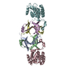
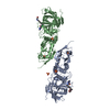
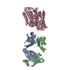
| Title | Crystal structure of the RV144 C1-C2 specific antibody CH54 Fab in complex with HIV-1 CLADE A/E GP120 and M48U1 | ||||||||||||
 Components Components |
| ||||||||||||
 Keywords Keywords | VIRAL PROTEIN/IMMUNE SYSTEM / ANTI-HIV-1 ENV ANTIBODY CH54 / CD4I ANTIBODY / ADCC / HIV-1 ENV / IMMUNE SYSTEM / RV144 VACCINE TRIAL / CLADE A/E 93TH057 / VIRAL PROTEIN-IMMUNE SYSTEM complex | ||||||||||||
| Function / homology |  Function and homology information Function and homology informationmembrane fusion involved in viral entry into host cell / viral envelope / symbiont entry into host cell / virion attachment to host cell / virion membrane Similarity search - Function | ||||||||||||
| Biological species |   Human immunodeficiency virus 1 Human immunodeficiency virus 1 Homo sapiens (human) Homo sapiens (human)synthetic construct (others) | ||||||||||||
| Method |  X-RAY DIFFRACTION / SYNCHROTRON / MOLECULAR REPLACEMENT / Resolution: 2.91 Å X-RAY DIFFRACTION / SYNCHROTRON / MOLECULAR REPLACEMENT / Resolution: 2.91 Å | ||||||||||||
 Authors Authors | Van, V. / Tolbert, W.D. / Pazgier, M. | ||||||||||||
| Funding support |  United States, 3items United States, 3items
| ||||||||||||
 Citation Citation | Journal: Mbio / Year: 2020 Title: Recognition Patterns of the C1/C2 Epitopes Involved in Fc-Mediated Response in HIV-1 Natural Infection and the RV114 Vaccine Trial. Authors: Tolbert, W.D. / Van, V. / Sherburn, R. / Tuyishime, M. / Yan, F. / Nguyen, D.N. / Stanfield-Oakley, S. / Easterhoff, D. / Bonsignori, M. / Haynes, B.F. / Moody, M.A. / Ray, K. / Ferrari, G. ...Authors: Tolbert, W.D. / Van, V. / Sherburn, R. / Tuyishime, M. / Yan, F. / Nguyen, D.N. / Stanfield-Oakley, S. / Easterhoff, D. / Bonsignori, M. / Haynes, B.F. / Moody, M.A. / Ray, K. / Ferrari, G. / Lewis, G.K. / Pazgier, M. | ||||||||||||
| History |
|
- Structure visualization
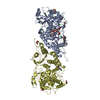
| Structure viewer | Molecule: MolmilJmol/JSmol |
|---|
- Downloads & links
-Download
| PDBx/mmCIF format | 6mg7.cif.gz | 318.1 KB | Display | PDBx/mmCIF format |
|---|---|---|---|---|
| PDB format | pdb6mg7.ent.gz | 257.7 KB | Display | PDB format |
| PDBx/mmJSON format | 6mg7.json.gz | Tree view | PDBx/mmJSON format | |
| Others |  Other downloads Other downloads |
-Validation report
| Arichive directory | https://data.pdbj.org/pub/pdb/validation_reports/mg/6mg7ftp://data.pdbj.org/pub/pdb/validation_reports/mg/6mg7 | HTTPS FTP |
|---|
-Related structure data
| Related structure data | 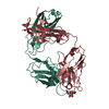 4fz8C 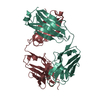 6oedC 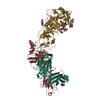 6oejC 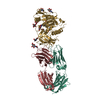 6ofiC  4rfoS S: Starting model for refinement C: citing same article ( |
|---|---|
| Similar structure data |


-Links
 PDBj
PDBj


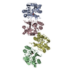
- Assembly
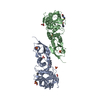
| Deposited unit | 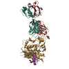
| ||||||||
|---|---|---|---|---|---|---|---|---|---|
| 1 |
| ||||||||
| Unit cell |
|
-Components
| #1: Protein | Mass: 42847.676 Da / Num. of mol.: 1 / Mutation: H375S Source method: isolated from a genetically manipulated source Source: (gene. exp.) Human immunodeficiency virus 1 / Gene: HIV-1 Env / Cell (production host): HEK 293 GNT1- cells / Production host: Homo sapiens (human) / References: UniProt: A0A0M3KKW9 | ||
|---|---|---|---|
| #2: Protein/peptide |   Type: Peptide-like / Class: Inhibitor / Mass: 3056.804 Da / Num. of mol.: 1 / Source method: obtained synthetically / Source: (synth.) synthetic construct (others) / References: CD4-MIMETIC MINIPROTEIN M48U1 Type: Peptide-like / Class: Inhibitor / Mass: 3056.804 Da / Num. of mol.: 1 / Source method: obtained synthetically / Source: (synth.) synthetic construct (others) / References: CD4-MIMETIC MINIPROTEIN M48U1 | ||
| #3: Antibody | Mass: 23532.475 Da / Num. of mol.: 1 Source method: isolated from a genetically manipulated source Source: (gene. exp.) Homo sapiens (human) / Cell (production host): HEK 293 / Production host: Homo sapiens (human) | ||
| #4: Antibody | Mass: 22637.182 Da / Num. of mol.: 1 Source method: isolated from a genetically manipulated source Source: (gene. exp.) Homo sapiens (human) / Cell (production host): HEK 293 / Production host: Homo sapiens (human) | ||
| #5: Sugar | ChemComp-NAG /   Type: D-saccharide, beta linking / Mass: 221.208 Da / Num. of mol.: 10 / Source method: obtained synthetically / Formula: C8H15NO6 Type: D-saccharide, beta linking / Mass: 221.208 Da / Num. of mol.: 10 / Source method: obtained synthetically / Formula: C8H15NO6Compound details | THE CD4-MIMETIC MINIPROTEINS INHIBIT HIV-1 ENTRY AND ARE DERIVED FROM SCYLLATOXIN (A SCORPION TOXIN) ...THE CD4-MIMETIC MINIPROTEI | |
-Experimental details
-Experiment
| Experiment | Method: X-RAY DIFFRACTION / Number of used crystals: 1 |
|---|
- Sample preparation
| Crystal | Density Matthews: 2.17 Å3/Da / Density % sol: 43.36 % |
|---|---|
| Crystal grow | Temperature: 294 K / Method: vapor diffusion, hanging drop / pH: 5.5 / Details: 25% PEG 4000 0.1 M MES pH 5.5 |
-Data collection
| Diffraction | Mean temperature: 100 K |
|---|---|
| Diffraction source | Source: SYNCHROTRON / Site: SSRL / Beamline: BL12-2 / Wavelength: 0.97946 Å |
| Detector | Type: DECTRIS PILATUS 6M / Detector: PIXEL / Date: Mar 25, 2018 |
| Radiation | Monochromator: Si (111) / Protocol: SINGLE WAVELENGTH / Monochromatic (M) / Laue (L): M / Scattering type: x-ray |
| Radiation wavelength | Wavelength: 0.97946 Å / Relative weight: 1 |
| Reflection | Resolution: 2.9→50 Å / Num. obs: 16963 / % possible obs: 95.9 % / Redundancy: 3.2 % / Rmerge(I) obs: 0.174 / Net I/σ(I): 11.1 |
| Reflection shell | Resolution: 2.9→2.95 Å / Redundancy: 3.1 % / Rmerge(I) obs: 0.833 / Mean I/σ(I) obs: 1 / % possible all: 97.3 |
- Processing
| Software |
| ||||||||||||||||||||||||||||||||||||||||||||||||||||||||||||||||||||||||||||||||||||||||||||||||||||||||||||||||||||||||||||||||||||||||||||||||||||||||||||||||||||||||||||||||||||||
|---|---|---|---|---|---|---|---|---|---|---|---|---|---|---|---|---|---|---|---|---|---|---|---|---|---|---|---|---|---|---|---|---|---|---|---|---|---|---|---|---|---|---|---|---|---|---|---|---|---|---|---|---|---|---|---|---|---|---|---|---|---|---|---|---|---|---|---|---|---|---|---|---|---|---|---|---|---|---|---|---|---|---|---|---|---|---|---|---|---|---|---|---|---|---|---|---|---|---|---|---|---|---|---|---|---|---|---|---|---|---|---|---|---|---|---|---|---|---|---|---|---|---|---|---|---|---|---|---|---|---|---|---|---|---|---|---|---|---|---|---|---|---|---|---|---|---|---|---|---|---|---|---|---|---|---|---|---|---|---|---|---|---|---|---|---|---|---|---|---|---|---|---|---|---|---|---|---|---|---|---|---|---|---|
| Refinement | Method to determine structure: MOLECULAR REPLACEMENT Starting model: 4RFO Resolution: 2.91→39.68 Å / Cor.coef. Fo:Fc: 0.918 / Cor.coef. Fo:Fc free: 0.872 / SU B: 85.574 / SU ML: 0.71 / Cross valid method: THROUGHOUT / ESU R Free: 0.571 / Details: HYDROGENS HAVE BEEN ADDED IN THE RIDING POSITIONS
| ||||||||||||||||||||||||||||||||||||||||||||||||||||||||||||||||||||||||||||||||||||||||||||||||||||||||||||||||||||||||||||||||||||||||||||||||||||||||||||||||||||||||||||||||||||||
| Solvent computation | Ion probe radii: 0.8 Å / Shrinkage radii: 0.8 Å / VDW probe radii: 1.2 Å | ||||||||||||||||||||||||||||||||||||||||||||||||||||||||||||||||||||||||||||||||||||||||||||||||||||||||||||||||||||||||||||||||||||||||||||||||||||||||||||||||||||||||||||||||||||||
| Displacement parameters | Biso mean: 89.89 Å2
| ||||||||||||||||||||||||||||||||||||||||||||||||||||||||||||||||||||||||||||||||||||||||||||||||||||||||||||||||||||||||||||||||||||||||||||||||||||||||||||||||||||||||||||||||||||||
| Refinement step | Cycle: LAST / Resolution: 2.91→39.68 Å
| ||||||||||||||||||||||||||||||||||||||||||||||||||||||||||||||||||||||||||||||||||||||||||||||||||||||||||||||||||||||||||||||||||||||||||||||||||||||||||||||||||||||||||||||||||||||
| Refine LS restraints |
|