 Movie
Movie Controller


+ Open data

- Basic information
| Entry | Database: PDB / ID: 4yve | ||||||
|---|---|---|---|---|---|---|---|
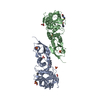
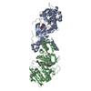
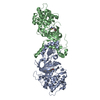
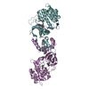
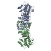
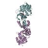
| Title | ROCK 1 bound to methoxyphenyl thiazole inhibitor | ||||||
 Components Components | Rho-associated protein kinase 1 | ||||||
 Keywords Keywords | TRANSFERASE/TRANSFERASE INHIBITOR / kinase / dimer / dimerization / myosin / TRANSFERASE-TRANSFERASE INHIBITOR complex | ||||||
| Function / homology |  Function and homology information Function and homology informationmyoblast migration / podocyte cell migration / apical constriction / regulation of angiotensin-activated signaling pathway / ROCK-subfamily protein kinase / membrane to membrane docking / regulation of keratinocyte differentiation / Rho-dependent protein serine/threonine kinase activity / negative regulation of membrane protein ectodomain proteolysis / positive regulation of connective tissue replacement ...myoblast migration / podocyte cell migration / apical constriction / regulation of angiotensin-activated signaling pathway / ROCK-subfamily protein kinase / membrane to membrane docking / regulation of keratinocyte differentiation / Rho-dependent protein serine/threonine kinase activity / negative regulation of membrane protein ectodomain proteolysis / positive regulation of connective tissue replacement / response to transforming growth factor beta / regulation of cell junction assembly / negative regulation of amyloid precursor protein catabolic process / embryonic morphogenesis / bleb assembly / regulation of cell motility / positive regulation of amyloid-beta clearance / leukocyte tethering or rolling / neuron projection arborization / bleb / negative regulation of biomineral tissue development / response to angiotensin / negative regulation of motor neuron apoptotic process / regulation of establishment of endothelial barrier / regulation of synapse maturation / regulation of stress fiber assembly / RHO GTPases Activate ROCKs / actomyosin structure organization / motor neuron apoptotic process / Sema4D induced cell migration and growth-cone collapse / cortical actin cytoskeleton organization / aortic valve morphogenesis / RND3 GTPase cycle / regulation of neuron differentiation / leukocyte migration / regulation of focal adhesion assembly / RHOBTB1 GTPase cycle / RHOB GTPase cycle / tau-protein kinase activity / leukocyte cell-cell adhesion / EPHA-mediated growth cone collapse / Apoptotic cleavage of cellular proteins / smooth muscle contraction / positive regulation of cardiac muscle hypertrophy / regulation of establishment of cell polarity / negative regulation of bicellular tight junction assembly / mRNA destabilization / RHOC GTPase cycle / negative regulation of amyloid-beta formation / mitotic cytokinesis / epithelial to mesenchymal transition / RHOH GTPase cycle / positive regulation of focal adhesion assembly / RHOA GTPase cycle / negative regulation of protein binding / regulation of synaptic vesicle endocytosis / regulation of cell adhesion / Rho protein signal transduction / ruffle / EPHB-mediated forward signaling / centriole / positive regulation of autophagy / regulation of cell migration / negative regulation of angiogenesis / blood vessel diameter maintenance / protein localization to plasma membrane / peptidyl-serine phosphorylation / regulation of microtubule cytoskeleton organization / regulation of actin cytoskeleton organization / VEGFA-VEGFR2 Pathway / tau protein binding / Schaffer collateral - CA1 synapse / small GTPase binding / neuron projection development / cytoplasmic stress granule / lamellipodium / G alpha (12/13) signalling events / actin cytoskeleton organization / secretory granule lumen / Potential therapeutics for SARS / cytoskeleton / positive regulation of MAPK cascade / intracellular signal transduction / Golgi membrane / protein serine kinase activity / protein serine/threonine kinase activity / positive regulation of gene expression / Neutrophil degranulation / signal transduction / extracellular region / zinc ion binding / ATP binding / plasma membrane / cytosol / cytoplasm Similarity search - Function | ||||||
| Biological species |  Homo sapiens (human) Homo sapiens (human) | ||||||
| Method |  X-RAY DIFFRACTION / SYNCHROTRON / FOURIER SYNTHESIS / Resolution: 3.4 Å X-RAY DIFFRACTION / SYNCHROTRON / FOURIER SYNTHESIS / Resolution: 3.4 Å | ||||||
 Authors Authors | Jacobs, M.D. | ||||||
 Citation Citation | Journal: J.Med.Chem. / Year: 2015 Title: Design, Synthesis, and Structure-Activity Relationships of Pyridine-Based Rho Kinase (ROCK) Inhibitors. Authors: Green, J. / Cao, J. / Bandarage, U.K. / Gao, H. / Court, J. / Marhefka, C. / Jacobs, M. / Taslimi, P. / Newsome, D. / Nakayama, T. / Shah, S. / Rodems, S. #1: Journal: J. Biol. Chem. / Year: 2006Title: The structure of dimeric ROCK I reveals the mechanism for ligand selectivity. Authors: Jacobs, M. / Hayakawa, K. / Swenson, L. / Bellon, S. / Fleming, M. / Taslimi, P. / Doran, J. #2: Journal: Bioorg. Med. Chem. Lett. / Year: 2018Title: ROCK inhibitors 2. Improving potency, selectivity and solubility through the application of rationally designed solubilizing groups. Authors: Gao, H. / Marhefka, C. / Jacobs, M.D. / Cao, J. / Bandarage, U.K. / Green, J. | ||||||
| History |
|
- Structure visualization
| Structure viewer | Molecule: MolmilJmol/JSmol |
|---|
- Downloads & links
-Download
| PDBx/mmCIF format | 4yve.cif.gz | 335.3 KB | Display | PDBx/mmCIF format |
|---|---|---|---|---|
| PDB format | pdb4yve.ent.gz | 278.5 KB | Display | PDB format |
| PDBx/mmJSON format | 4yve.json.gz | Tree view | PDBx/mmJSON format | |
| Others |  Other downloads Other downloads |
-Validation report
| Arichive directory | https://data.pdbj.org/pub/pdb/validation_reports/yv/4yveftp://data.pdbj.org/pub/pdb/validation_reports/yv/4yve | HTTPS FTP |
|---|
-Related structure data
| Related structure data |  4yvcC  5bmlC  2etrS C: citing same article ( S: Starting model for refinement |
|---|---|
| Similar structure data |


-Links
 PDBj
PDBj


- Assembly
| Deposited unit | 
| ||||||||
|---|---|---|---|---|---|---|---|---|---|
| 1 |
| ||||||||
| Unit cell |
|
-Components
| #1: Protein | Mass: 48012.285 Da / Num. of mol.: 2 / Fragment: N-terminal kinase domain (UNP residues 6-415) Source method: isolated from a genetically manipulated source Source: (gene. exp.) Homo sapiens (human) / Gene: ROCK1 / Plasmid: pBEV10 / Production host:   Spodoptera frugiperda (fall armyworm) Spodoptera frugiperda (fall armyworm)References: UniProt: Q13464, non-specific serine/threonine protein kinase #2: Chemical |   Mass: 325.385 Da / Num. of mol.: 2 / Source method: obtained synthetically / Formula: C17H15N3O2S Mass: 325.385 Da / Num. of mol.: 2 / Source method: obtained synthetically / Formula: C17H15N3O2S#3: Water | ChemComp-HOH / |  Mass: 18.015 Da / Num. of mol.: 9 / Source method: isolated from a natural source / Formula: H2O Mass: 18.015 Da / Num. of mol.: 9 / Source method: isolated from a natural source / Formula: H2O |
|---|
-Experimental details
-Experiment
| Experiment | Method: X-RAY DIFFRACTION / Number of used crystals: 1 |
|---|
- Sample preparation
| Crystal | Density Matthews: 4.51 Å3/Da / Density % sol: 72.71 % |
|---|---|
| Crystal grow | Temperature: 298 K / Method: vapor diffusion, hanging drop / pH: 5.5 Details: 0.45 mM protein, 5% PEG3350, 100 mM MES, 50 mM calcium chloride, 10 mM DTT |
-Data collection
| Diffraction | Mean temperature: 100 K | |||||||||||||||||||||||||||||||||||||||||||||||||||||||||||||||||||||||||||||||||||||||||||||||||||
|---|---|---|---|---|---|---|---|---|---|---|---|---|---|---|---|---|---|---|---|---|---|---|---|---|---|---|---|---|---|---|---|---|---|---|---|---|---|---|---|---|---|---|---|---|---|---|---|---|---|---|---|---|---|---|---|---|---|---|---|---|---|---|---|---|---|---|---|---|---|---|---|---|---|---|---|---|---|---|---|---|---|---|---|---|---|---|---|---|---|---|---|---|---|---|---|---|---|---|---|---|
| Diffraction source | Source: SYNCHROTRON / Site: ALS  / Beamline: 5.0.2 / Wavelength: 1.1 Å / Beamline: 5.0.2 / Wavelength: 1.1 Å | |||||||||||||||||||||||||||||||||||||||||||||||||||||||||||||||||||||||||||||||||||||||||||||||||||
| Detector | Type: ADSC QUANTUM 4 / Detector: CCD / Date: Jan 28, 2003 | |||||||||||||||||||||||||||||||||||||||||||||||||||||||||||||||||||||||||||||||||||||||||||||||||||
| Radiation | Monochromator: double crystal Si(111) / Protocol: SINGLE WAVELENGTH / Monochromatic (M) / Laue (L): M / Scattering type: x-ray | |||||||||||||||||||||||||||||||||||||||||||||||||||||||||||||||||||||||||||||||||||||||||||||||||||
| Radiation wavelength | Wavelength: 1.1 Å / Relative weight: 1 | |||||||||||||||||||||||||||||||||||||||||||||||||||||||||||||||||||||||||||||||||||||||||||||||||||
| Reflection | Resolution: 3.4→29.78 Å / Num. obs: 23765 / % possible obs: 99 % / Redundancy: 7.41 % / Biso Wilson estimate: 94.4 Å2 / Rmerge(I) obs: 0.155 / Χ2: 2.29 / Net I/σ(I): 4.2 / Num. measured all: 178283 / Scaling rejects: 2109 | |||||||||||||||||||||||||||||||||||||||||||||||||||||||||||||||||||||||||||||||||||||||||||||||||||
| Reflection shell | Diffraction-ID: 1
|
- Processing
| Software |
| |||||||||||||||||||||||||||||||||||||||||||||||||||||||||||||||||||||||||||||||||||||||||||||||||||||||||||||||||||||||||||||||||||||||||||||||||||||||||||||||||||||||||||||||||||||||||||||||||||||||||||||||||||||||||||||||||
|---|---|---|---|---|---|---|---|---|---|---|---|---|---|---|---|---|---|---|---|---|---|---|---|---|---|---|---|---|---|---|---|---|---|---|---|---|---|---|---|---|---|---|---|---|---|---|---|---|---|---|---|---|---|---|---|---|---|---|---|---|---|---|---|---|---|---|---|---|---|---|---|---|---|---|---|---|---|---|---|---|---|---|---|---|---|---|---|---|---|---|---|---|---|---|---|---|---|---|---|---|---|---|---|---|---|---|---|---|---|---|---|---|---|---|---|---|---|---|---|---|---|---|---|---|---|---|---|---|---|---|---|---|---|---|---|---|---|---|---|---|---|---|---|---|---|---|---|---|---|---|---|---|---|---|---|---|---|---|---|---|---|---|---|---|---|---|---|---|---|---|---|---|---|---|---|---|---|---|---|---|---|---|---|---|---|---|---|---|---|---|---|---|---|---|---|---|---|---|---|---|---|---|---|---|---|---|---|---|---|---|---|---|---|---|---|---|---|---|---|---|---|---|---|---|---|---|
| Refinement | Method to determine structure: FOURIER SYNTHESIS Starting model: PDB entry 2ETR Resolution: 3.4→19.86 Å / Cor.coef. Fo:Fc: 0.9051 / Cor.coef. Fo:Fc free: 0.8771 / Cross valid method: THROUGHOUT / σ(F): 0 / SU Rfree Blow DPI: 0.427
| |||||||||||||||||||||||||||||||||||||||||||||||||||||||||||||||||||||||||||||||||||||||||||||||||||||||||||||||||||||||||||||||||||||||||||||||||||||||||||||||||||||||||||||||||||||||||||||||||||||||||||||||||||||||||||||||||
| Displacement parameters | Biso max: 184.88 Å2 / Biso mean: 85.88 Å2 / Biso min: 20 Å2
| |||||||||||||||||||||||||||||||||||||||||||||||||||||||||||||||||||||||||||||||||||||||||||||||||||||||||||||||||||||||||||||||||||||||||||||||||||||||||||||||||||||||||||||||||||||||||||||||||||||||||||||||||||||||||||||||||
| Refine analyze | Luzzati coordinate error obs: 0.728 Å | |||||||||||||||||||||||||||||||||||||||||||||||||||||||||||||||||||||||||||||||||||||||||||||||||||||||||||||||||||||||||||||||||||||||||||||||||||||||||||||||||||||||||||||||||||||||||||||||||||||||||||||||||||||||||||||||||
| Refinement step | Cycle: final / Resolution: 3.4→19.86 Å
| |||||||||||||||||||||||||||||||||||||||||||||||||||||||||||||||||||||||||||||||||||||||||||||||||||||||||||||||||||||||||||||||||||||||||||||||||||||||||||||||||||||||||||||||||||||||||||||||||||||||||||||||||||||||||||||||||
| Refine LS restraints |
| |||||||||||||||||||||||||||||||||||||||||||||||||||||||||||||||||||||||||||||||||||||||||||||||||||||||||||||||||||||||||||||||||||||||||||||||||||||||||||||||||||||||||||||||||||||||||||||||||||||||||||||||||||||||||||||||||
| LS refinement shell | Resolution: 3.4→3.55 Å / Total num. of bins used: 12
| |||||||||||||||||||||||||||||||||||||||||||||||||||||||||||||||||||||||||||||||||||||||||||||||||||||||||||||||||||||||||||||||||||||||||||||||||||||||||||||||||||||||||||||||||||||||||||||||||||||||||||||||||||||||||||||||||
| Refinement TLS params. | Method: refined / Refine-ID: X-RAY DIFFRACTION
| |||||||||||||||||||||||||||||||||||||||||||||||||||||||||||||||||||||||||||||||||||||||||||||||||||||||||||||||||||||||||||||||||||||||||||||||||||||||||||||||||||||||||||||||||||||||||||||||||||||||||||||||||||||||||||||||||
| Refinement TLS group |
|
