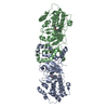
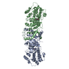
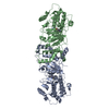
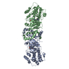
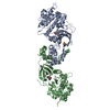
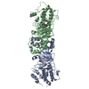
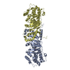
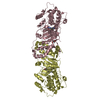
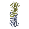
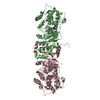
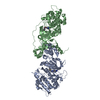
Entry Database : PDB / ID : 2etrTitle Crystal Structure of ROCK I bound to Y-27632 Rho-associated protein kinase 1 Keywords / / / / / Function / homology Function Domain/homology Component
/ / / / / / / / / / / / / / / / / / / / / / / / / / / / / / / / / / / / / / / / / / / / / / / / / / / / / / / / / / / / / / / / / / / / / / / / / / / / / / / / / / / / / / / / / / / / / / / / / / / / / / / / / / / / / / / / / / / / / / / / / / / / / / / / / / / / / / / / / / / / / / / / / / / / / Biological species Homo sapiens (human)Method / / / Resolution : 2.6 Å Authors Jacobs, M. Journal : J.Biol.Chem. / Year : 2006Title : The Structure of Dimeric ROCK I Reveals the Mechanism for Ligand Selectivity.Authors : Jacobs, M. / Hayakawa, K. / Swenson, L. / Bellon, S. / Fleming, M. / Taslimi, P. / Doran, J. History Deposition Oct 27, 2005 Deposition site / Processing site Revision 1.0 Nov 8, 2005 Provider / Type Revision 1.1 May 1, 2008 Group Revision 1.2 Jul 13, 2011 Group Revision 1.3 Oct 18, 2017 Group / Category Revision 1.4 Aug 23, 2023 Group Data collection / Database references ... Data collection / Database references / Derived calculations / Refinement description Category chem_comp_atom / chem_comp_bond ... chem_comp_atom / chem_comp_bond / database_2 / pdbx_initial_refinement_model / struct_ref_seq_dif / struct_site Item _database_2.pdbx_DOI / _database_2.pdbx_database_accession ... _database_2.pdbx_DOI / _database_2.pdbx_database_accession / _struct_ref_seq_dif.details / _struct_site.pdbx_auth_asym_id / _struct_site.pdbx_auth_comp_id / _struct_site.pdbx_auth_seq_id
Show all Show less
 Movie
Movie Controller


 Open data
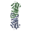
 Basic information
 Components
 Keywords
 Function and homology information
 Homo sapiens (human)
 X-RAY DIFFRACTION /
  Authors
 Citation
 Structure visualization
 Downloads & links
 Other downloads


 PDBj
PDBj


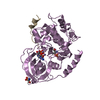
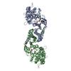
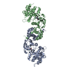
 Assembly
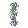

 Spodoptera frugiperda (fall armyworm) / References: UniProt: Q13464, EC: 2.7.1.37
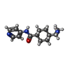
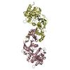
 Mass: 247.336 Da / Num. of mol.: 2 / Source method: obtained synthetically / Formula: C14H21N3O
 Mass: 18.015 Da / Num. of mol.: 108 / Source method: isolated from a natural source / Formula: H2O
 Sample preparation
 / Beamline: 32-ID / Wavelength: 1 Å
 Processing