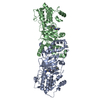
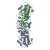
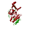
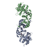
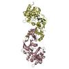
Entry Database : PDB / ID : 3nczTitle X-Ray Co-structure of Rho-Associated Protein Kinase (ROCK1) with a potent 2H-isoquinolin-1-one inhibitor Rho-associated protein kinase 1 Keywords / / / / / Function / homology Function Domain/homology Component
/ / / / / / / / / / / / / / / / / / / / / / / / / / / / / / / / / / / / / / / / / / / / / / / / / / / / / / / / / / / / / / / / / / / / / / / / / / / / / / / / / / / / / / / / / / / / / / / / / / / / / / / / / / / / / / / / / / / / / / / / / / / / / / / / / / / / / / / / / / / / / / / / / / / / / Biological species Homo sapiens (human)Method / / / Resolution : 3 Å Authors Li, X. Journal : Bioorg.Med.Chem.Lett. / Year : 2010Title : Substituted 2H-isoquinolin-1-ones as potent Rho-kinase inhibitors: Part 2, optimization for blood pressure reduction in spontaneously hypertensive rats.Authors : Ginn, J.D. / Bosanac, T. / Chen, R. / Cywin, C. / Hickey, E. / Kashem, M. / Kerr, S. / Kugler, S. / Li, X. / Prokopowicz, A. / Schlyer, S. / Smith, J.D. / Turner, M.R. / Wu, F. / Young, E.R. History Deposition Jun 6, 2010 Deposition site / Processing site Revision 1.0 Dec 8, 2010 Provider / Type Revision 1.1 Jul 13, 2011 Group Revision 1.2 Jul 17, 2019 Group / Refinement description / Category Item / _software.name / _software.versionRevision 1.3 Feb 21, 2024 Group / Database references / Derived calculationsCategory chem_comp_atom / chem_comp_bond ... chem_comp_atom / chem_comp_bond / database_2 / struct_ref_seq_dif / struct_site Item _database_2.pdbx_DOI / _database_2.pdbx_database_accession ... _database_2.pdbx_DOI / _database_2.pdbx_database_accession / _struct_ref_seq_dif.details / _struct_site.pdbx_auth_asym_id / _struct_site.pdbx_auth_comp_id / _struct_site.pdbx_auth_seq_id
Show all Show less
 Movie
Movie Controller

 Yorodumi
 Open data
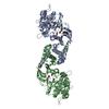
 Basic information
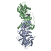
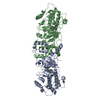
 Components
 Keywords
 Function and homology information
 Homo sapiens (human)
 X-RAY DIFFRACTION /
  Authors
 Citation
 Structure visualization
 Downloads & links
 Other downloads
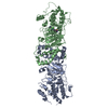


 PDBj
PDBj


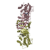
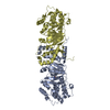
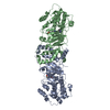
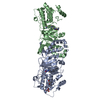
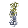
 Assembly
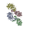

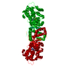
 Spodoptera frugiperda (fall armyworm)
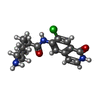
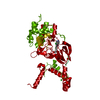
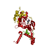
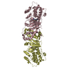
 Mass: 319.786 Da / Num. of mol.: 4 / Source method: obtained synthetically / Formula: C16H18ClN3O2
 Mass: 18.015 Da / Num. of mol.: 63 / Source method: isolated from a natural source / Formula: H2O
 Sample preparation
 / Beamline: X06DA / Wavelength: 0.8 Å
 Processing