 Movie
Movie Controller

[English] 日本語
 Yorodumi
- PDB-2erz: Crystal Structure of c-AMP Dependent Kinase (PKA) bound to hydrox... -
+ Open data

- Basic information
| Entry | Database: PDB / ID: 2erz | ||||||
|---|---|---|---|---|---|---|---|
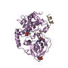
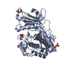
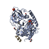
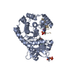
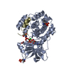
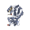
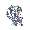
| Title | Crystal Structure of c-AMP Dependent Kinase (PKA) bound to hydroxyfasudil | ||||||
 Components Components |
| ||||||
 Keywords Keywords | TRANSFERASE / fasudil kinase | ||||||
| Function / homology |  Function and homology information Function and homology informationspontaneous exocytosis of neurotransmitter / PKA activation in glucagon signalling / CREB1 phosphorylation through the activation of Adenylate Cyclase / negative regulation of meiotic cell cycle / HDL assembly / positive regulation of triglyceride catabolic process / DARPP-32 events / Rap1 signalling / PKA activation / Regulation of insulin secretion ...spontaneous exocytosis of neurotransmitter / PKA activation in glucagon signalling / CREB1 phosphorylation through the activation of Adenylate Cyclase / negative regulation of meiotic cell cycle / HDL assembly / positive regulation of triglyceride catabolic process / DARPP-32 events / Rap1 signalling / PKA activation / Regulation of insulin secretion / Vasopressin regulates renal water homeostasis via Aquaporins / GPER1 signaling / Glucagon-like Peptide-1 (GLP1) regulates insulin secretion / Hedgehog 'off' state / Loss of Nlp from mitotic centrosomes / Recruitment of mitotic centrosome proteins and complexes / Loss of proteins required for interphase microtubule organization from the centrosome / Anchoring of the basal body to the plasma membrane / Recruitment of NuMA to mitotic centrosomes / AURKA Activation by TPX2 / Factors involved in megakaryocyte development and platelet production / MAPK6/MAPK4 signaling / Interleukin-3, Interleukin-5 and GM-CSF signaling / CD209 (DC-SIGN) signaling / RET signaling / GLI3 is processed to GLI3R by the proteasome / High laminar flow shear stress activates signaling by PIEZO1 and PECAM1:CDH5:KDR in endothelial cells / regulation of cellular respiration / Regulation of PLK1 Activity at G2/M Transition / Mitochondrial protein degradation / VEGFA-VEGFR2 Pathway / nucleotide-activated protein kinase complex / Ion homeostasis / cAMP-dependent protein kinase inhibitor activity / potassium channel inhibitor activity / histone H1-4S35 kinase activity / cAMP-dependent protein kinase / negative regulation of glycolytic process / regulation of protein processing / cAMP-dependent protein kinase activity / cellular response to parathyroid hormone stimulus / protein localization to lipid droplet / molecular function inhibitor activity / regulation of bicellular tight junction assembly / cAMP-dependent protein kinase complex / interleukin-2-mediated signaling pathway / sperm capacitation / regulation of osteoblast differentiation / peptidyl-threonine phosphorylation / cellular response to cold / protein kinase A regulatory subunit binding / negative regulation of glycolytic process through fructose-6-phosphate / ciliary base / protein kinase A catalytic subunit binding / intracellular potassium ion homeostasis / TORC1 signaling / mesoderm formation / plasma membrane raft / cAMP/PKA signal transduction / axoneme / sperm flagellum / postsynaptic modulation of chemical synaptic transmission / negative regulation of cAMP/PKA signal transduction / regulation of synaptic transmission, glutamatergic / negative regulation of TORC1 signaling / protein serine/threonine/tyrosine kinase activity / negative regulation of protein localization to chromatin / lipid droplet / cellular response to glucagon stimulus / cellular response to nutrient levels / positive regulation of gluconeogenesis / positive regulation of phagocytosis / acrosomal vesicle / regulation of proteasomal protein catabolic process / positive regulation of protein export from nucleus / neural tube closure / peptidyl-serine phosphorylation / regulation of microtubule cytoskeleton organization / cellular response to glucose stimulus / negative regulation of smoothened signaling pathway / neuromuscular junction / positive regulation of cholesterol biosynthetic process / modulation of chemical synaptic transmission / small GTPase binding / mRNA processing / adenylate cyclase-inhibiting G protein-coupled receptor signaling pathway / protein autophosphorylation / manganese ion binding / cellular response to heat / sperm midpiece / presynapse / adenylate cyclase-activating G protein-coupled receptor signaling pathway / molecular adaptor activity / protein phosphorylation / protein kinase activity / postsynapse / regulation of cell cycle / nuclear speck / protein domain specific binding / protein serine kinase activity Similarity search - Function | ||||||
| Biological species |  | ||||||
| Method |  X-RAY DIFFRACTION / MOLECULAR REPLACEMENT / Resolution: 2.2 Å X-RAY DIFFRACTION / MOLECULAR REPLACEMENT / Resolution: 2.2 Å | ||||||
 Authors Authors | Jacobs, M. | ||||||
 Citation Citation | Journal: J.Biol.Chem. / Year: 2006 Title: The Structure of Dimeric ROCK I Reveals the Mechanism for Ligand Selectivity. Authors: Jacobs, M. / Hayakawa, K. / Swenson, L. / Bellon, S. / Fleming, M. / Taslimi, P. / Doran, J. | ||||||
| History |
|
- Structure visualization
| Structure viewer | Molecule: MolmilJmol/JSmol |
|---|
- Downloads & links
-Download
| PDBx/mmCIF format | 2erz.cif.gz | 88.8 KB | Display | PDBx/mmCIF format |
|---|---|---|---|---|
| PDB format | pdb2erz.ent.gz | 65.7 KB | Display | PDB format |
| PDBx/mmJSON format | 2erz.json.gz | Tree view | PDBx/mmJSON format | |
| Others |  Other downloads Other downloads |
-Validation report
| Arichive directory | https://data.pdbj.org/pub/pdb/validation_reports/er/2erzftp://data.pdbj.org/pub/pdb/validation_reports/er/2erz | HTTPS FTP |
|---|
-Related structure data
| Related structure data | 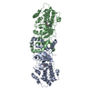 2esmC 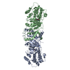 2etkC 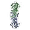 2etrC 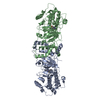 3d9vC 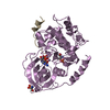 1atpS C: citing same article ( S: Starting model for refinement |
|---|---|
| Similar structure data |


-Links
 PDBj
PDBj


- Assembly
| Deposited unit | 
| ||||||||
|---|---|---|---|---|---|---|---|---|---|
| 1 |
| ||||||||
| Unit cell |
| ||||||||
| Details | Biological assembly is same as asym. unit: one protein molecule (PKA), one inhibitor peptide (PKI), one ligand (hydroxyfasudil) |
-Components
| #1: Protein | Mass: 40788.512 Da / Num. of mol.: 1 Source method: isolated from a genetically manipulated source Source: (gene. exp.)  |
|---|---|
| #2: Protein/peptide | Mass: 2226.411 Da / Num. of mol.: 1 / Fragment: residues 5-24 / Source method: obtained synthetically Details: This sequence occurs naturally in Oryctolagus cuniculus (rabbit) References: UniProt: P61926 |
| #3: Chemical | ChemComp-HFS /   Mass: 307.368 Da / Num. of mol.: 1 / Source method: obtained synthetically / Formula: C14H17N3O3S Mass: 307.368 Da / Num. of mol.: 1 / Source method: obtained synthetically / Formula: C14H17N3O3S |
| #4: Water | ChemComp-HOH /  Mass: 18.015 Da / Num. of mol.: 61 / Source method: isolated from a natural source / Formula: H2O Mass: 18.015 Da / Num. of mol.: 61 / Source method: isolated from a natural source / Formula: H2O |
| Has protein modification | Y |
-Experimental details
-Experiment
| Experiment | Method: X-RAY DIFFRACTION / Number of used crystals: 1 |
|---|
- Sample preparation
| Crystal | Density Matthews: 2.54 Å3/Da / Density % sol: 51.67 % |
|---|---|
| Crystal grow | Temperature: 277 K / Method: vapor diffusion, hanging drop / pH: 7 Details: bicine, pH 7.0, 2% MPD, VAPOR DIFFUSION, HANGING DROP, temperature 277K |
-Data collection
| Diffraction | Mean temperature: 110 K |
|---|---|
| Diffraction source | Source: ROTATING ANODE / Type: RIGAKU RU300 / Wavelength: 1.5418 Å |
| Detector | Type: RIGAKU RAXIS IV / Detector: IMAGE PLATE / Date: Mar 19, 2004 / Details: Osmic |
| Radiation | Monochromator: Osmic mirrors / Protocol: SINGLE WAVELENGTH / Monochromatic (M) / Laue (L): M / Scattering type: x-ray |
| Radiation wavelength | Wavelength: 1.5418 Å / Relative weight: 1 |
| Reflection | Resolution: 2.2→27.51 Å / Num. all: 23077 / Num. obs: 22293 / % possible obs: 96.6 % / Observed criterion σ(F): 1 / Observed criterion σ(I): 1 / Redundancy: 4.87 % / Rmerge(I) obs: 0.075 / Χ2: 0.99 / Net I/σ(I): 13.6 / Scaling rejects: 821 |
| Reflection shell | Resolution: 2.2→2.28 Å / % possible obs: 84.5 % / Rmerge(I) obs: 0.344 / Mean I/σ(I) obs: 3.4 / Num. measured obs: 263 / Χ2: 1.16 / % possible all: 96.6 |
- Processing
| Software |
| ||||||||||||||||||||||||||||||||||||
|---|---|---|---|---|---|---|---|---|---|---|---|---|---|---|---|---|---|---|---|---|---|---|---|---|---|---|---|---|---|---|---|---|---|---|---|---|---|
| Refinement | Method to determine structure: MOLECULAR REPLACEMENT Starting model: PDB ENTRY 1ATP Resolution: 2.2→19.8 Å / Rfactor Rfree error: 0.007 / Data cutoff high absF: 139000.516 / Data cutoff low absF: 0 / Isotropic thermal model: RESTRAINED / Cross valid method: THROUGHOUT / σ(F): 0 / Stereochemistry target values: Engh & Huber
| ||||||||||||||||||||||||||||||||||||
| Solvent computation | Solvent model: FLAT MODEL / Bsol: 44.029 Å2 / ksol: 0.327 e/Å3 | ||||||||||||||||||||||||||||||||||||
| Displacement parameters | Biso mean: 38 Å2
| ||||||||||||||||||||||||||||||||||||
| Refine analyze |
| ||||||||||||||||||||||||||||||||||||
| Refinement step | Cycle: LAST / Resolution: 2.2→19.8 Å
| ||||||||||||||||||||||||||||||||||||
| Refine LS restraints |
| ||||||||||||||||||||||||||||||||||||
| LS refinement shell | Resolution: 2.2→2.34 Å / Rfactor Rfree error: 0.024 / Total num. of bins used: 6
| ||||||||||||||||||||||||||||||||||||
| Xplor file |
|
