 Movie
Movie Controller

[English] 日本語
 Yorodumi
- PDB-1fmo: CRYSTAL STRUCTURE OF A POLYHISTIDINE-TAGGED RECOMBINANT CATALYTIC... -
+ Open data

- Basic information
| Entry | Database: PDB / ID: 1fmo | ||||||
|---|---|---|---|---|---|---|---|
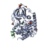
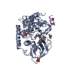
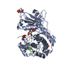
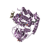
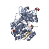
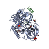
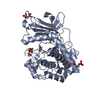
| Title | CRYSTAL STRUCTURE OF A POLYHISTIDINE-TAGGED RECOMBINANT CATALYTIC SUBUNIT OF CAMP-DEPENDENT PROTEIN KINASE COMPLEXED WITH THE PEPTIDE INHIBITOR PKI(5-24) AND ADENOSINE | ||||||
 Components Components |
| ||||||
 Keywords Keywords | COMPLEX (PHOSPHOTRANSFERASE/INHIBITOR) / COMPLEX (PHOSPHOTRANSFERASE-INHIBITOR) / PHOSPHORYLATION / POLYHISTIDINE-TAG / PROTEIN KINASE / COMPLEX (PHOSPHOTRANSFERASE-INHIBITOR) complex | ||||||
| Function / homology |  Function and homology information Function and homology informationspontaneous exocytosis of neurotransmitter / PKA activation in glucagon signalling / CREB1 phosphorylation through the activation of Adenylate Cyclase / negative regulation of meiotic cell cycle / HDL assembly / positive regulation of triglyceride catabolic process / DARPP-32 events / Rap1 signalling / PKA activation / Regulation of insulin secretion ...spontaneous exocytosis of neurotransmitter / PKA activation in glucagon signalling / CREB1 phosphorylation through the activation of Adenylate Cyclase / negative regulation of meiotic cell cycle / HDL assembly / positive regulation of triglyceride catabolic process / DARPP-32 events / Rap1 signalling / PKA activation / Regulation of insulin secretion / Vasopressin regulates renal water homeostasis via Aquaporins / GPER1 signaling / Glucagon-like Peptide-1 (GLP1) regulates insulin secretion / Hedgehog 'off' state / Loss of Nlp from mitotic centrosomes / Recruitment of mitotic centrosome proteins and complexes / Loss of proteins required for interphase microtubule organization from the centrosome / Anchoring of the basal body to the plasma membrane / Recruitment of NuMA to mitotic centrosomes / AURKA Activation by TPX2 / Factors involved in megakaryocyte development and platelet production / MAPK6/MAPK4 signaling / Interleukin-3, Interleukin-5 and GM-CSF signaling / CD209 (DC-SIGN) signaling / RET signaling / GLI3 is processed to GLI3R by the proteasome / High laminar flow shear stress activates signaling by PIEZO1 and PECAM1:CDH5:KDR in endothelial cells / Regulation of PLK1 Activity at G2/M Transition / Mitochondrial protein degradation / VEGFA-VEGFR2 Pathway / nucleotide-activated protein kinase complex / Ion homeostasis / regulation of cellular respiration / cAMP-dependent protein kinase inhibitor activity / histone H1-4S35 kinase activity / cAMP-dependent protein kinase / regulation of protein processing / negative regulation of glycolytic process / cAMP-dependent protein kinase activity / cellular response to parathyroid hormone stimulus / protein localization to lipid droplet / molecular function inhibitor activity / regulation of bicellular tight junction assembly / cAMP-dependent protein kinase complex / negative regulation of protein import into nucleus / interleukin-2-mediated signaling pathway / regulation of osteoblast differentiation / sperm capacitation / cellular response to cold / protein kinase A regulatory subunit binding / negative regulation of glycolytic process through fructose-6-phosphate / ciliary base / protein kinase A catalytic subunit binding / intracellular potassium ion homeostasis / mesoderm formation / TORC1 signaling / plasma membrane raft / cAMP/PKA signal transduction / axoneme / sperm flagellum / postsynaptic modulation of chemical synaptic transmission / negative regulation of cAMP/PKA signal transduction / regulation of synaptic transmission, glutamatergic / regulation of G2/M transition of mitotic cell cycle / negative regulation of TORC1 signaling / protein serine/threonine/tyrosine kinase activity / regulation of proteasomal protein catabolic process / lipid droplet / cellular response to glucagon stimulus / cellular response to nutrient levels / positive regulation of phagocytosis / positive regulation of gluconeogenesis / acrosomal vesicle / positive regulation of protein export from nucleus / neuromuscular junction / negative regulation of smoothened signaling pathway / neural tube closure / cellular response to glucose stimulus / positive regulation of cholesterol biosynthetic process / modulation of chemical synaptic transmission / small GTPase binding / mRNA processing / adenylate cyclase-inhibiting G protein-coupled receptor signaling pathway / manganese ion binding / cellular response to heat / adenylate cyclase-activating G protein-coupled receptor signaling pathway / presynapse / sperm midpiece / molecular adaptor activity / protein kinase activity / regulation of cell cycle / nuclear speck / postsynapse / protein domain specific binding / protein serine kinase activity / protein serine/threonine kinase activity / positive regulation of cell population proliferation / ubiquitin protein ligase binding / centrosome / protein kinase binding Similarity search - Function | ||||||
| Biological species |  | ||||||
| Method |  X-RAY DIFFRACTION / ISOMORPHOUS REPLACEMENT / Resolution: 2.2 Å X-RAY DIFFRACTION / ISOMORPHOUS REPLACEMENT / Resolution: 2.2 Å | ||||||
 Authors Authors | Narayana, N. / Cox, S. / Shaltiel, S. / Taylor, S.S. / Xuong, N.-H. | ||||||
 Citation Citation | Journal: Biochemistry / Year: 1997 Title: Crystal structure of a polyhistidine-tagged recombinant catalytic subunit of cAMP-dependent protein kinase complexed with the peptide inhibitor PKI(5-24) and adenosine. Authors: Narayana, N. / Cox, S. / Shaltiel, S. / Taylor, S.S. / Xuong, N. #1: Journal: Structure / Year: 1997Title: A Binary Complex of the Catalytic Subunit of Camp-Dependent Protein Kinase and Adenosine Further Defines Conformational Flexibility Authors: Narayana, N. / Cox, S. / Xuong, N.H. / Ten Eyck, L.F. / Taylor, S.S. #2: Journal: Biochemistry / Year: 1993Title: Crystal Structure of the Catalytic Subunit of Camp-Dependent Protein Kinase Complexed with Mgatp and Peptide Inhibitor Authors: Zheng, J. / Knighton, D.R. / Ten Eyck, L.F. / Karlsson, R. / Xuong, N.H. / Taylor, S.S. / Sowadski, J.M. #3: Journal: Protein Eng. / Year: 1993Title: Expression of the Catalytic Subunit of Camp-Dependent Protein Kinase in Escherichia Coli: Multiple Isozymes Reflect Different Phosphorylation States Authors: Herberg, F.W. / Bell, S.M. / Taylor, S.S. #4: Journal: Science / Year: 1991Title: Crystal Structure of the Catalytic Subunit of Cyclic Adenosine Monophosphate-Dependent Protein Kinase Authors: Knighton, D.R. / Zheng, J.H. / Ten Eyck, L.F. / Ashford, V.A. / Xuong, N.H. / Taylor, S.S. / Sowadski, J.M. #5: Journal: Science / Year: 1991Title: Structure of a Peptide Inhibitor Bound to the Catalytic Subunit of Cyclic Adenosine Monophosphate-Dependent Protein Kinase Authors: Knighton, D.R. / Zheng, J.H. / Ten Eyck, L.F. / Xuong, N.H. / Taylor, S.S. / Sowadski, J.M. | ||||||
| History |
|
- Structure visualization
| Structure viewer | Molecule: MolmilJmol/JSmol |
|---|
- Downloads & links
-Download
| PDBx/mmCIF format | 1fmo.cif.gz | 90.6 KB | Display | PDBx/mmCIF format |
|---|---|---|---|---|
| PDB format | pdb1fmo.ent.gz | 67 KB | Display | PDB format |
| PDBx/mmJSON format | 1fmo.json.gz | Tree view | PDBx/mmJSON format | |
| Others |  Other downloads Other downloads |
-Validation report
| Arichive directory | https://data.pdbj.org/pub/pdb/validation_reports/fm/1fmoftp://data.pdbj.org/pub/pdb/validation_reports/fm/1fmo | HTTPS FTP |
|---|
-Related structure data
| Related structure data |  1apmS S: Starting model for refinement |
|---|---|
| Similar structure data |


-Links
 PDBj
PDBj


- Assembly
| Deposited unit | 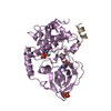
| ||||||||
|---|---|---|---|---|---|---|---|---|---|
| 1 |
| ||||||||
| Unit cell |
|
-Components
| #1: Protein | Mass: 40657.316 Da / Num. of mol.: 1 / Fragment: CATALYTIC SUBUNIT Source method: isolated from a genetically manipulated source Source: (gene. exp.)  |
|---|---|
| #2: Protein/peptide | Mass: 2226.411 Da / Num. of mol.: 1 / Fragment: RESIDUES 5 - 24 Source method: isolated from a genetically manipulated source References: UniProt: P61926 |
| #3: Chemical | ChemComp-ADN /   Mass: 267.241 Da / Num. of mol.: 1 / Source method: obtained synthetically / Formula: C10H13N5O4 Mass: 267.241 Da / Num. of mol.: 1 / Source method: obtained synthetically / Formula: C10H13N5O4 |
| #4: Water | ChemComp-HOH /  Mass: 18.015 Da / Num. of mol.: 90 / Source method: isolated from a natural source / Formula: H2O Mass: 18.015 Da / Num. of mol.: 90 / Source method: isolated from a natural source / Formula: H2O |
| Has protein modification | Y |
-Experimental details
-Experiment
| Experiment | Method: X-RAY DIFFRACTION / Number of used crystals: 1 |
|---|
- Sample preparation
| Crystal | Density Matthews: 2.7 Å3/Da / Density % sol: 54 % | ||||||||||||||||||||||||||||||||||||||||
|---|---|---|---|---|---|---|---|---|---|---|---|---|---|---|---|---|---|---|---|---|---|---|---|---|---|---|---|---|---|---|---|---|---|---|---|---|---|---|---|---|---|
| Crystal grow | Temperature: 277 K / Method: vapor diffusion, hanging drop / pH: 8 Details: ORTHORHOMBIC CRYSTALS OF THE TERNARY COMPLEX, COMPRISED OF MOUSE HIS6-RC SUBUNIT, PKI (5-24) AND ADENOSINE, WERE GROWN BY THE HANGING-DROP VAPOUR DIFFUSION METHOD USING 2-METHYL-2,4- ...Details: ORTHORHOMBIC CRYSTALS OF THE TERNARY COMPLEX, COMPRISED OF MOUSE HIS6-RC SUBUNIT, PKI (5-24) AND ADENOSINE, WERE GROWN BY THE HANGING-DROP VAPOUR DIFFUSION METHOD USING 2-METHYL-2,4-PENTANEDIOL (MPD) AS THE PRECIPITATING AGENT. THE MOTHER-LIQUOR CONTAINED PROTEIN AT A CONCENTRATION OF 0.25 MM IN 100 MM BICINE BUFFER AT PH 8.0, 0.75 MM PKI(5-24), 4 MM ADENOSINE AND 4% MPD. THE RESERVOIR SOLUTION WAS MADE UP OF 20% MPD IN 100 MM BICINE BUFFER (PH 8.0). X-RAY DIFFRACTION QUALITY CRYSTALS (0.2 X 0.15 X 0.8 MM3) WERE GROWN AT 4 C IN APPROXIMATELY 4 - 6 WEEKS., vapor diffusion - hanging drop, temperature 277K | ||||||||||||||||||||||||||||||||||||||||
| Crystal grow | *PLUS Temperature: 4 ℃ / Method: vapor diffusion, hanging drop | ||||||||||||||||||||||||||||||||||||||||
| Components of the solutions | *PLUS
|
-Data collection
| Diffraction | Mean temperature: 277 K |
|---|---|
| Diffraction source | Source: ROTATING ANODE / Type: RIGAKU RUH2R / Wavelength: 1.5418 |
| Detector | Type: XUONG-HAMLIN MULTIWIRE / Detector: AREA DETECTOR / Date: Feb 7, 1994 |
| Radiation | Monochromator: GRAPHITE(002) / Monochromatic (M) / Laue (L): M / Scattering type: x-ray |
| Radiation wavelength | Wavelength: 1.5418 Å / Relative weight: 1 |
| Reflection | Resolution: 2.2→20 Å / Num. obs: 21679 / % possible obs: 92 % / Observed criterion σ(I): 0 / Redundancy: 2.4 % / Biso Wilson estimate: 24 Å2 / Rsym value: 0.064 / Net I/σ(I): 7.3 |
| Reflection shell | Resolution: 2.2→2.3 Å / Redundancy: 1.4 % / Mean I/σ(I) obs: 1 / Rsym value: 0.25 / % possible all: 84 |
| Reflection | *PLUS Rmerge(I) obs: 0.064 |
| Reflection shell | *PLUS % possible obs: 84 % / Rmerge(I) obs: 0.25 |
- Processing
| Software |
| ||||||||||||||||||||||||||||||||||||||||||||||||||
|---|---|---|---|---|---|---|---|---|---|---|---|---|---|---|---|---|---|---|---|---|---|---|---|---|---|---|---|---|---|---|---|---|---|---|---|---|---|---|---|---|---|---|---|---|---|---|---|---|---|---|---|
| Refinement | Method to determine structure: ISOMORPHOUS REPLACEMENT Starting model: PDB ENTRY 1APM Resolution: 2.2→10 Å / σ(F): 1
| ||||||||||||||||||||||||||||||||||||||||||||||||||
| Solvent computation | Bsol: 167.1 Å2 / ksol: 0.45 e/Å3 | ||||||||||||||||||||||||||||||||||||||||||||||||||
| Refinement step | Cycle: LAST / Resolution: 2.2→10 Å
| ||||||||||||||||||||||||||||||||||||||||||||||||||
| Refine LS restraints |
| ||||||||||||||||||||||||||||||||||||||||||||||||||
| Software | *PLUS Name: TNT / Version: 5E / Classification: refinement | ||||||||||||||||||||||||||||||||||||||||||||||||||
| Refinement | *PLUS Rfactor obs: 0.182 | ||||||||||||||||||||||||||||||||||||||||||||||||||
| Solvent computation | *PLUS | ||||||||||||||||||||||||||||||||||||||||||||||||||
| Displacement parameters | *PLUS | ||||||||||||||||||||||||||||||||||||||||||||||||||
| Refine LS restraints | *PLUS
|
