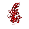
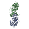
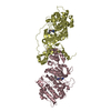
Entry Database : PDB / ID : 3v8sTitle Human RHO-ASSOCIATED PROTEIN KINASE 1 (ROCK 1) IN COMPLEX WITH INDAZOLE DERIVATIVE (COMPOUND 18) Rho-associated protein kinase 1 Keywords / / / / / / Function / homology Function Domain/homology Component
/ / / / / / / / / / / / / / / / / / / / / / / / / / / / / / / / / / / / / / / / / / / / / / / / / / / / / / / / / / / / / / / / / / / / / / / / / / / / / / / / / / / / / / / / / / / / / / / / / / / / / / / / / / / / / / / / / / / / / / / / / / / / / / / / / / / / / / / / / / / / / / / / / / Biological species Homo sapiens (human)Method / / / Resolution : 2.286 Å Authors Martin, M.P. / Zhu, J.-Yi. / Schonbrunn, E. Journal : J.Med.Chem. / Year : 2012Title : Fragment-based and structure-guided discovery and optimization of rho kinase inhibitors.Authors : Li, R. / Martin, M.P. / Liu, Y. / Wang, B. / Patel, R.A. / Zhu, J.Y. / Sun, N. / Pireddu, R. / Lawrence, N.J. / Li, J. / Haura, E.B. / Sung, S.S. / Guida, W.C. / Schonbrunn, E. / Sebti, S.M. History Deposition Dec 23, 2011 Deposition site / Processing site Revision 1.0 Feb 8, 2012 Provider / Type Revision 1.1 Mar 21, 2012 Group Revision 1.2 Sep 13, 2023 Group Data collection / Database references ... Data collection / Database references / Derived calculations / Refinement description Category chem_comp_atom / chem_comp_bond ... chem_comp_atom / chem_comp_bond / database_2 / pdbx_initial_refinement_model / struct_site Item _database_2.pdbx_DOI / _database_2.pdbx_database_accession ... _database_2.pdbx_DOI / _database_2.pdbx_database_accession / _struct_site.pdbx_auth_asym_id / _struct_site.pdbx_auth_comp_id / _struct_site.pdbx_auth_seq_id
Show all Show less
 Movie
Movie Controller

 Yorodumi
 Open data
 Basic information
 Components
 Keywords
 Function and homology information
 Homo sapiens (human)
 X-RAY DIFFRACTION /
  Authors
 Citation
 Structure visualization
 Downloads & links
 Other downloads


 PDBj
PDBj


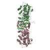
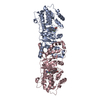
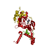
 Assembly
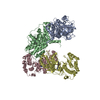

 Spodoptera frugiperda (fall armyworm)
 Mass: 280.324 Da / Num. of mol.: 4 / Source method: obtained synthetically / Formula: C16H16N4O
 Mass: 18.015 Da / Num. of mol.: 872 / Source method: isolated from a natural source / Formula: H2O
 Sample preparation
 / Beamline: 22-ID / Wavelength: 1 Å
 Processing