 Movie
Movie Controller

[English] 日本語
 Yorodumi
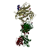
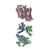
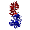
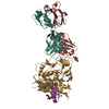
- PDB-4ye4: Crystal Structure of Neutralizing Antibody HJ16 in Complex with H... -
+ Open data

- Basic information
| Entry | Database: PDB / ID: 4ye4 | ||||||
|---|---|---|---|---|---|---|---|
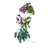
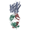
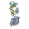
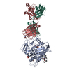
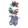
| Title | Crystal Structure of Neutralizing Antibody HJ16 in Complex with HIV-1 gp120 | ||||||
 Components Components |
| ||||||
 Keywords Keywords | Viral protein/Immune system / HIV1 / antibody / gp120 / complex / Viral protein-Immune system complex | ||||||
| Function / homology |  Function and homology information Function and homology information: / host cell endosome membrane / clathrin-dependent endocytosis of virus by host cell / fusion of virus membrane with host endosome membrane / viral envelope / virion attachment to host cell / host cell plasma membrane / virion membrane / structural molecule activity / membrane Similarity search - Function | ||||||
| Biological species |   Human immunodeficiency virus Human immunodeficiency virus Homo sapiens (human) Homo sapiens (human) | ||||||
| Method |  X-RAY DIFFRACTION / SYNCHROTRON / MOLECULAR REPLACEMENT / molecular replacement / Resolution: 2.72 Å X-RAY DIFFRACTION / SYNCHROTRON / MOLECULAR REPLACEMENT / molecular replacement / Resolution: 2.72 Å | ||||||
 Authors Authors | Kwong, P.D. / Chen, L. / Zhou, T. | ||||||
| Funding support |  United States, 1items United States, 1items
| ||||||
 Citation Citation | Journal: Cell / Year: 2015 Title: Structural Repertoire of HIV-1-Neutralizing Antibodies Targeting the CD4 Supersite in 14 Donors. Authors: Zhou, T. / Lynch, R.M. / Chen, L. / Acharya, P. / Wu, X. / Doria-Rose, N.A. / Joyce, M.G. / Lingwood, D. / Soto, C. / Bailer, R.T. / Ernandes, M.J. / Kong, R. / Longo, N.S. / Louder, M.K. / ...Authors: Zhou, T. / Lynch, R.M. / Chen, L. / Acharya, P. / Wu, X. / Doria-Rose, N.A. / Joyce, M.G. / Lingwood, D. / Soto, C. / Bailer, R.T. / Ernandes, M.J. / Kong, R. / Longo, N.S. / Louder, M.K. / McKee, K. / O'Dell, S. / Schmidt, S.D. / Tran, L. / Yang, Z. / Druz, A. / Luongo, T.S. / Moquin, S. / Srivatsan, S. / Yang, Y. / Zhang, B. / Zheng, A. / Pancera, M. / Kirys, T. / Georgiev, I.S. / Gindin, T. / Peng, H.P. / Yang, A.S. / Mullikin, J.C. / Gray, M.D. / Stamatatos, L. / Burton, D.R. / Koff, W.C. / Cohen, M.S. / Haynes, B.F. / Casazza, J.P. / Connors, M. / Corti, D. / Lanzavecchia, A. / Sattentau, Q.J. / Weiss, R.A. / West Jr., A.P. / Bjorkman, P.J. / Scheid, J.F. / Nussenzweig, M.C. / Shapiro, L. / Mascola, J.R. / Kwong, P.D. | ||||||
| History |
|
- Structure visualization
| Structure viewer | Molecule: MolmilJmol/JSmol |
|---|
- Downloads & links
-Download
| PDBx/mmCIF format | 4ye4.cif.gz | 321.2 KB | Display | PDBx/mmCIF format |
|---|---|---|---|---|
| PDB format | pdb4ye4.ent.gz | 259.8 KB | Display | PDB format |
| PDBx/mmJSON format | 4ye4.json.gz | Tree view | PDBx/mmJSON format | |
| Others |  Other downloads Other downloads |
-Validation report
| Arichive directory | https://data.pdbj.org/pub/pdb/validation_reports/ye/4ye4ftp://data.pdbj.org/pub/pdb/validation_reports/ye/4ye4 | HTTPS FTP |
|---|
-Related structure data
| Related structure data |  4rwyC 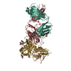 4rx4C 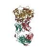 4ydiC 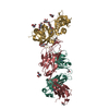 4ydjC 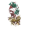 4ydkC 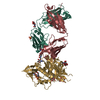 4ydlC 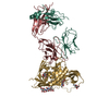 4yflC C: citing same article ( |
|---|---|
| Similar structure data |

-Links
 PDBj
PDBj


- Assembly
| Deposited unit | 
| ||||||||
|---|---|---|---|---|---|---|---|---|---|
| 1 |
| ||||||||
| Unit cell |
|
-Components
| #1: Protein | Mass: 39492.484 Da / Num. of mol.: 1 Source method: isolated from a genetically manipulated source Details: HIV-1 clade B gp120 core / Source: (gene. exp.) Human immunodeficiency virus / Production host: Homo sapiens (human) / References: UniProt: Q69994*PLUS | ||||
|---|---|---|---|---|---|
| #2: Antibody | Mass: 24606.832 Da / Num. of mol.: 1 Source method: isolated from a genetically manipulated source Source: (gene. exp.) Homo sapiens (human) / Production host: Homo sapiens (human) | ||||
| #3: Antibody | Mass: 24202.906 Da / Num. of mol.: 1 Source method: isolated from a genetically manipulated source Source: (gene. exp.) Homo sapiens (human) / Production host: Homo sapiens (human) | ||||
| #4: Sugar | ChemComp-NAG /   Type: D-saccharide, beta linking / Mass: 221.208 Da / Num. of mol.: 8 Type: D-saccharide, beta linking / Mass: 221.208 Da / Num. of mol.: 8Source method: isolated from a genetically manipulated source Formula: C8H15NO6 #5: Water | ChemComp-HOH / |  Mass: 18.015 Da / Num. of mol.: 27 / Source method: isolated from a natural source / Formula: H2O Mass: 18.015 Da / Num. of mol.: 27 / Source method: isolated from a natural source / Formula: H2OHas protein modification | Y | |
-Experimental details
-Experiment
| Experiment | Method: X-RAY DIFFRACTION / Number of used crystals: 1 |
|---|
- Sample preparation
| Crystal | Density Matthews: 2.23 Å3/Da / Density % sol: 44.82 % |
|---|---|
| Crystal grow | Temperature: 293 K / Method: vapor diffusion, hanging drop Details: 0.1 M imidazole pH 6.5 10 % (W/V) PEG 8000 5% isopropanol PH range: 6-7 |
-Data collection
| Diffraction | Mean temperature: 200 K |
|---|---|
| Diffraction source | Source: SYNCHROTRON / Site: APS / Beamline: 22-ID / Wavelength: 1 Å |
| Detector | Type: MARMOSAIC 300 mm CCD / Detector: CCD / Date: Apr 14, 2013 |
| Radiation | Monochromator: Si 220 / Protocol: SINGLE WAVELENGTH / Monochromatic (M) / Laue (L): M / Scattering type: x-ray |
| Radiation wavelength | Wavelength: 1 Å / Relative weight: 1 |
| Reflection | Resolution: 2.72→41.9 Å / Num. obs: 20510 / % possible obs: 94.4 % / Redundancy: 4.6 % / Net I/σ(I): 1.98 |
| Reflection shell | Highest resolution: 2.72 Å |
-Phasing
| Phasing | Method: molecular replacement |
|---|
- Processing
| Software |
| |||||||||||||||||||||||||||||||||||||||||||||||||||||||||||||||||||||||||||||||||||||||||||||||||||||||||||||||||||||||||||||||||||||||||||||||||||||||||||||||||||||||||||||||
|---|---|---|---|---|---|---|---|---|---|---|---|---|---|---|---|---|---|---|---|---|---|---|---|---|---|---|---|---|---|---|---|---|---|---|---|---|---|---|---|---|---|---|---|---|---|---|---|---|---|---|---|---|---|---|---|---|---|---|---|---|---|---|---|---|---|---|---|---|---|---|---|---|---|---|---|---|---|---|---|---|---|---|---|---|---|---|---|---|---|---|---|---|---|---|---|---|---|---|---|---|---|---|---|---|---|---|---|---|---|---|---|---|---|---|---|---|---|---|---|---|---|---|---|---|---|---|---|---|---|---|---|---|---|---|---|---|---|---|---|---|---|---|---|---|---|---|---|---|---|---|---|---|---|---|---|---|---|---|---|---|---|---|---|---|---|---|---|---|---|---|---|---|---|---|---|---|
| Refinement | Method to determine structure: MOLECULAR REPLACEMENT / Resolution: 2.72→41.9 Å / SU ML: 0.36 / Cross valid method: FREE R-VALUE / σ(F): 1.34 / Phase error: 26.33 / Stereochemistry target values: ML
| |||||||||||||||||||||||||||||||||||||||||||||||||||||||||||||||||||||||||||||||||||||||||||||||||||||||||||||||||||||||||||||||||||||||||||||||||||||||||||||||||||||||||||||||
| Solvent computation | Shrinkage radii: 0.9 Å / VDW probe radii: 1.11 Å / Solvent model: FLAT BULK SOLVENT MODEL | |||||||||||||||||||||||||||||||||||||||||||||||||||||||||||||||||||||||||||||||||||||||||||||||||||||||||||||||||||||||||||||||||||||||||||||||||||||||||||||||||||||||||||||||
| Displacement parameters | Biso max: 186.81 Å2 / Biso mean: 60.9245 Å2 / Biso min: 20 Å2 | |||||||||||||||||||||||||||||||||||||||||||||||||||||||||||||||||||||||||||||||||||||||||||||||||||||||||||||||||||||||||||||||||||||||||||||||||||||||||||||||||||||||||||||||
| Refinement step | Cycle: final / Resolution: 2.72→41.9 Å
| |||||||||||||||||||||||||||||||||||||||||||||||||||||||||||||||||||||||||||||||||||||||||||||||||||||||||||||||||||||||||||||||||||||||||||||||||||||||||||||||||||||||||||||||
| LS refinement shell | Highest resolution: 2.72 Å | |||||||||||||||||||||||||||||||||||||||||||||||||||||||||||||||||||||||||||||||||||||||||||||||||||||||||||||||||||||||||||||||||||||||||||||||||||||||||||||||||||||||||||||||
| Refinement TLS params. | Method: refined / Refine-ID: X-RAY DIFFRACTION
| |||||||||||||||||||||||||||||||||||||||||||||||||||||||||||||||||||||||||||||||||||||||||||||||||||||||||||||||||||||||||||||||||||||||||||||||||||||||||||||||||||||||||||||||
| Refinement TLS group |
|
