 Movie
Movie Controller

[English] 日本語
 Yorodumi
- PDB-6ob0: Compound 2 bound structure of WT Lipoprotein Lipase in Complex wi... -
+ Open data

- Basic information
| Entry | Database: PDB / ID: 6ob0 | ||||||
|---|---|---|---|---|---|---|---|
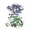
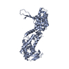
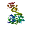
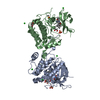
| Title | Compound 2 bound structure of WT Lipoprotein Lipase in Complex with GPIHBP1 Mutant N78D N82D produced in HEK293-F cells | ||||||
 Components Components |
| ||||||
 Keywords Keywords | HYDROLASE/PROTEIN BINDING / Lipase / HYDROLASE-PROTEIN BINDING complex | ||||||
| Function / homology |  Function and homology information Function and homology informationchylomicron binding / positive regulation of chylomicron remodeling / positive regulation of chylomicron remnant clearance / lipoprotein lipase / lipoprotein lipase activity / lipoprotein lipase activator activity / low-density lipoprotein particle mediated signaling / chylomicron remodeling / positive regulation of cholesterol storage / response to heparin ...chylomicron binding / positive regulation of chylomicron remodeling / positive regulation of chylomicron remnant clearance / lipoprotein lipase / lipoprotein lipase activity / lipoprotein lipase activator activity / low-density lipoprotein particle mediated signaling / chylomicron remodeling / positive regulation of cholesterol storage / response to heparin / phospholipase A1 / glycerophospholipase activity / lipase binding / glycerophospholipid phospholipase A1 activity / Assembly of active LPL and LIPC lipase complexes / Post-translational modification: synthesis of GPI-anchored proteins / triglyceride catabolic process / protein transporter activity / very-low-density lipoprotein particle remodeling / transcytosis / Chylomicron remodeling / very-low-density lipoprotein particle clearance / chylomicron / high-density lipoprotein particle remodeling / positive regulation of fatty acid biosynthetic process / positive regulation of lipid storage / cellular response to nutrient / protein import / triacylglycerol lipase activity / very-low-density lipoprotein particle / positive regulation of macrophage derived foam cell differentiation / cellular response to fatty acid / heparan sulfate proteoglycan binding / positive regulation of chemokine (C-X-C motif) ligand 2 production / protein localization to cell surface / triglyceride metabolic process / triglyceride homeostasis / lipoprotein particle binding / apolipoprotein binding / positive regulation of fat cell differentiation / retinoid metabolic process / response to glucose / Retinoid metabolism and transport / catalytic complex / positive regulation of chemokine production / phospholipid metabolic process / protein-membrane adaptor activity / positive regulation of adipose tissue development / cholesterol homeostasis / positive regulation of interleukin-1 beta production / response to bacterium / fatty acid metabolic process / intracellular protein transport / Transcriptional regulation of white adipocyte differentiation / positive regulation of interleukin-6 production / fatty acid biosynthetic process / positive regulation of tumor necrosis factor production / positive regulation of inflammatory response / heparin binding / MLL4 and MLL3 complexes regulate expression of PPARG target genes in adipogenesis and hepatic steatosis / basolateral plasma membrane / apical plasma membrane / protein stabilization / signaling receptor binding / external side of plasma membrane / calcium ion binding / lipid binding / cell surface / protein homodimerization activity / : / extracellular region / plasma membrane Similarity search - Function | ||||||
| Biological species |  Homo sapiens (human) Homo sapiens (human) | ||||||
| Method |  X-RAY DIFFRACTION / SYNCHROTRON / MOLECULAR REPLACEMENT / Resolution: 2.81 Å X-RAY DIFFRACTION / SYNCHROTRON / MOLECULAR REPLACEMENT / Resolution: 2.81 Å | ||||||
 Authors Authors | Arora, R. / Horton, P.A. / Benson, T.E. / Romanowski, M.J. | ||||||
 Citation Citation | Journal: Proc.Natl.Acad.Sci.USA / Year: 2019 Title: Structure of lipoprotein lipase in complex with GPIHBP1. Authors: Arora, R. / Nimonkar, A.V. / Baird, D. / Wang, C. / Chiu, C.H. / Horton, P.A. / Hanrahan, S. / Cubbon, R. / Weldon, S. / Tschantz, W.R. / Mueller, S. / Brunner, R. / Lehr, P. / Meier, P. / ...Authors: Arora, R. / Nimonkar, A.V. / Baird, D. / Wang, C. / Chiu, C.H. / Horton, P.A. / Hanrahan, S. / Cubbon, R. / Weldon, S. / Tschantz, W.R. / Mueller, S. / Brunner, R. / Lehr, P. / Meier, P. / Ottl, J. / Voznesensky, A. / Pandey, P. / Smith, T.M. / Stojanovic, A. / Flyer, A. / Benson, T.E. / Romanowski, M.J. / Trauger, J.W. | ||||||
| History |
|
- Structure visualization
| Structure viewer | Molecule: MolmilJmol/JSmol |
|---|
- Downloads & links
-Download
| PDBx/mmCIF format | 6ob0.cif.gz | 433.8 KB | Display | PDBx/mmCIF format |
|---|---|---|---|---|
| PDB format | pdb6ob0.ent.gz | 354.2 KB | Display | PDB format |
| PDBx/mmJSON format | 6ob0.json.gz | Tree view | PDBx/mmJSON format | |
| Others |  Other downloads Other downloads |
-Validation report
| Arichive directory | https://data.pdbj.org/pub/pdb/validation_reports/ob/6ob0ftp://data.pdbj.org/pub/pdb/validation_reports/ob/6ob0 | HTTPS FTP |
|---|
-Related structure data
| Related structure data |  6oauC  6oazSC S: Starting model for refinement C: citing same article ( |
|---|---|
| Similar structure data |


-Links
 PDBj
PDBj


- Assembly
| Deposited unit | 
| ||||||||
|---|---|---|---|---|---|---|---|---|---|
| 1 | 
| ||||||||
| 2 | 
| ||||||||
| 3 | 
| ||||||||
| 4 | 
| ||||||||
| Unit cell |
|
-Components
-Protein , 2 types, 8 molecules ABCDEFGH
| #1: Protein | Mass: 50465.133 Da / Num. of mol.: 4 Source method: isolated from a genetically manipulated source Source: (gene. exp.) Homo sapiens (human) / Gene: GPIHBP1, HBP1 / Cell line (production host): HEK293-F / Production host: Homo sapiens (human) / References: UniProt: P06858, lipoprotein lipase#2: Protein | Mass: 14727.757 Da / Num. of mol.: 4 / Fragment: residues 21-151 / Mutation: N78D, N82D Source method: isolated from a genetically manipulated source Source: (gene. exp.) Homo sapiens (human) / Gene: GPIHBP1, HBP1 / Cell line (production host): HEK293-F / Production host: Homo sapiens (human) / References: UniProt: Q8IV16 |
|---|
-Sugars , 1 types, 8 molecules 
| #3: Sugar | ChemComp-NAG /  Type: D-saccharide, beta linking / Mass: 221.208 Da / Num. of mol.: 8 Type: D-saccharide, beta linking / Mass: 221.208 Da / Num. of mol.: 8Source method: isolated from a genetically manipulated source Formula: C8H15NO6 |
|---|
-Non-polymers , 5 types, 247 molecules 


| #4: Chemical | ChemComp-M3D /  Mass: 467.516 Da / Num. of mol.: 8 / Source method: obtained synthetically / Formula: C28H25N3O4 / Feature type: SUBJECT OF INVESTIGATION Mass: 467.516 Da / Num. of mol.: 8 / Source method: obtained synthetically / Formula: C28H25N3O4 / Feature type: SUBJECT OF INVESTIGATION#5: Chemical | ChemComp-EDO /  Mass: 62.068 Da / Num. of mol.: 8 / Source method: obtained synthetically / Formula: C2H6O2 Mass: 62.068 Da / Num. of mol.: 8 / Source method: obtained synthetically / Formula: C2H6O2#6: Chemical | ChemComp-CA /  Mass: 40.078 Da / Num. of mol.: 4 / Source method: obtained synthetically / Formula: Ca Mass: 40.078 Da / Num. of mol.: 4 / Source method: obtained synthetically / Formula: Ca#7: Chemical | ChemComp-TRS / |  Mass: 122.143 Da / Num. of mol.: 1 / Source method: obtained synthetically / Formula: C4H12NO3 / Comment: pH buffer*YM Mass: 122.143 Da / Num. of mol.: 1 / Source method: obtained synthetically / Formula: C4H12NO3 / Comment: pH buffer*YM#8: Water | ChemComp-HOH / | Mass: 18.015 Da / Num. of mol.: 226 / Source method: isolated from a natural source / Formula: H2O |
|---|
-Details
| Has protein modification | Y |
|---|
-Experimental details
-Experiment
| Experiment | Method: X-RAY DIFFRACTION / Number of used crystals: 1 |
|---|
- Sample preparation
| Crystal | Density Matthews: 2.74 Å3/Da / Density % sol: 55.05 % |
|---|---|
| Crystal grow | Temperature: 298 K / Method: vapor diffusion, sitting drop / Details: 0.15M calcium acetate, 18% PEG3350 |
-Data collection
| Diffraction | Mean temperature: 100 K / Serial crystal experiment: N | ||||||||||||||||||||||||
|---|---|---|---|---|---|---|---|---|---|---|---|---|---|---|---|---|---|---|---|---|---|---|---|---|---|
| Diffraction source | Source: SYNCHROTRON / Site: APS  / Beamline: 17-ID / Wavelength: 1 Å / Beamline: 17-ID / Wavelength: 1 Å | ||||||||||||||||||||||||
| Detector | Type: DECTRIS PILATUS 6M / Detector: PIXEL / Date: Mar 7, 2018 | ||||||||||||||||||||||||
| Radiation | Protocol: SINGLE WAVELENGTH / Monochromatic (M) / Laue (L): M / Scattering type: x-ray | ||||||||||||||||||||||||
| Radiation wavelength | Wavelength: 1 Å / Relative weight: 1 | ||||||||||||||||||||||||
| Reflection | Resolution: 2.81→37.1 Å / Num. obs: 70015 / % possible obs: 99.1 % / Redundancy: 6.4 % / CC1/2: 0.998 / Rmerge(I) obs: 0.148 / Rpim(I) all: 0.064 / Rrim(I) all: 0.162 / Net I/σ(I): 12 | ||||||||||||||||||||||||
| Reflection shell | Diffraction-ID: 1
|
- Processing
| Software |
| ||||||||||||||||||||||||||||||||||||||||||||||||||||||||||||||||||||||||||||||||||||||||||||||||||||||||||||
|---|---|---|---|---|---|---|---|---|---|---|---|---|---|---|---|---|---|---|---|---|---|---|---|---|---|---|---|---|---|---|---|---|---|---|---|---|---|---|---|---|---|---|---|---|---|---|---|---|---|---|---|---|---|---|---|---|---|---|---|---|---|---|---|---|---|---|---|---|---|---|---|---|---|---|---|---|---|---|---|---|---|---|---|---|---|---|---|---|---|---|---|---|---|---|---|---|---|---|---|---|---|---|---|---|---|---|---|---|---|
| Refinement | Method to determine structure: MOLECULAR REPLACEMENT Starting model: 6OAZ Resolution: 2.81→37.15 Å / Cor.coef. Fo:Fc: 0.903 / Cor.coef. Fo:Fc free: 0.891 / SU R Cruickshank DPI: 2.176 / Cross valid method: THROUGHOUT / σ(F): 0 / SU R Blow DPI: 1.927 / SU Rfree Blow DPI: 0.319 / SU Rfree Cruickshank DPI: 0.325
| ||||||||||||||||||||||||||||||||||||||||||||||||||||||||||||||||||||||||||||||||||||||||||||||||||||||||||||
| Displacement parameters | Biso max: 172.97 Å2 / Biso mean: 67.92 Å2 / Biso min: 29.32 Å2
| ||||||||||||||||||||||||||||||||||||||||||||||||||||||||||||||||||||||||||||||||||||||||||||||||||||||||||||
| Refine analyze | Luzzati coordinate error obs: 0.36 Å | ||||||||||||||||||||||||||||||||||||||||||||||||||||||||||||||||||||||||||||||||||||||||||||||||||||||||||||
| Refinement step | Cycle: final / Resolution: 2.81→37.15 Å
| ||||||||||||||||||||||||||||||||||||||||||||||||||||||||||||||||||||||||||||||||||||||||||||||||||||||||||||
| Refine LS restraints |
| ||||||||||||||||||||||||||||||||||||||||||||||||||||||||||||||||||||||||||||||||||||||||||||||||||||||||||||
| LS refinement shell | Resolution: 2.81→2.83 Å / Rfactor Rfree error: 0 / Total num. of bins used: 50
|
