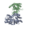
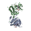
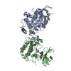
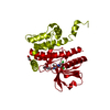
positive regulation of sodium ion import across plasma membrane / negative regulation of pancreatic juice secretion / negative regulation of sodium ion transmembrane transport / negative regulation of transmembrane transport / chemokine (C-X-C motif) ligand 12 signaling pathway / maintenance of lens transparency / monoatomic ion homeostasis / negative regulation of potassium ion transmembrane transport / positive regulation of potassium ion import across plasma membrane / intracellular chloride ion homeostasis ...positive regulation of sodium ion import across plasma membrane / negative regulation of pancreatic juice secretion / negative regulation of sodium ion transmembrane transport / negative regulation of transmembrane transport / chemokine (C-X-C motif) ligand 12 signaling pathway / maintenance of lens transparency / monoatomic ion homeostasis / negative regulation of potassium ion transmembrane transport / positive regulation of potassium ion import across plasma membrane / intracellular chloride ion homeostasis / positive regulation of T cell chemotaxis / renal sodium ion absorption / cellular hypotonic response / macrophage activation / positive regulation of potassium ion transport / cellular response to chemokine / cellular hyperosmotic response / ion channel regulator activity / response to aldosterone / potassium channel inhibitor activity / positive regulation of p38MAPK cascade / cellular response to potassium ion / cell volume homeostasis / regulation of monoatomic cation transmembrane transport / peptidyl-threonine phosphorylation / response to dietary excess / transporter activator activity / potassium ion transmembrane transport / sodium ion transmembrane transport / regulation of blood pressure / kinase activity / protein autophosphorylation / cell body / regulation of inflammatory response / cell cortex / basolateral plasma membrane / protein phosphorylation / transmembrane transporter binding / protein kinase activity / non-specific serine/threonine protein kinase / apical plasma membrane / intracellular signal transduction / inflammatory response / protein serine kinase activity / protein serine/threonine kinase activity / protein kinase binding / signal transduction / nucleoplasm / ATP binding / membrane / cytoplasm / cytosol Similarity search - Function
Serine/threonine-protein kinase OSR1/WNK, CCT domain / Oxidative-stress-responsive kinase 1 C-terminal domain / : / Phosphorylase Kinase; domain 1 / Phosphorylase Kinase; domain 1 / Transferase(Phosphotransferase) domain 1 / Transferase(Phosphotransferase); domain 1 / Protein kinase domain / Serine/Threonine protein kinases, catalytic domain / Protein kinase, ATP binding site ...Serine/threonine-protein kinase OSR1/WNK, CCT domain / Oxidative-stress-responsive kinase 1 C-terminal domain / : / Phosphorylase Kinase; domain 1 / Phosphorylase Kinase; domain 1 / Transferase(Phosphotransferase) domain 1 / Transferase(Phosphotransferase); domain 1 / Protein kinase domain / Serine/Threonine protein kinases, catalytic domain / Protein kinase, ATP binding site / Protein kinases ATP-binding region signature. / Protein kinase domain profile. / Protein kinase domain / Protein kinase-like domain superfamily / 2-Layer Sandwich / Orthogonal Bundle / Mainly Alpha / Alpha Beta Similarity search - Domain/homology
In the structure databanks used in Yorodumi, some data are registered as the other names, "COVID-19 virus" and "2019-nCoV". Here are the details of the virus and the list of structure data.
Jan 31, 2019. EMDB accession codes are about to change! (news from PDBe EMDB page)
EMDB accession codes are about to change! (news from PDBe EMDB page)
The allocation of 4 digits for EMDB accession codes will soon come to an end. Whilst these codes will remain in use, new EMDB accession codes will include an additional digit and will expand incrementally as the available range of codes is exhausted. The current 4-digit format prefixed with “EMD-” (i.e. EMD-XXXX) will advance to a 5-digit format (i.e. EMD-XXXXX), and so on. It is currently estimated that the 4-digit codes will be depleted around Spring 2019, at which point the 5-digit format will come into force.
The EM Navigator/Yorodumi systems omit the EMD- prefix.
Related info.:Q: What is EMD? / ID/Accession-code notation in Yorodumi/EM Navigator
Yorodumi is a browser for structure data from EMDB, PDB, SASBDB, etc.
This page is also the successor to EM Navigator detail page, and also detail information page/front-end page for Omokage search.
The word "yorodu" (or yorozu) is an old Japanese word meaning "ten thousand". "mi" (miru) is to see.
Related info.:EMDB / PDB / SASBDB / Comparison of 3 databanks / Yorodumi Search / Aug 31, 2016. New EM Navigator & Yorodumi / Yorodumi Papers / Jmol/JSmol / Function and homology information / Changes in new EM Navigator and Yorodumi
 Movie
Movie Controller

 Yorodumi
 Open data
 Basic information
 Components
 Keywords
 Function and homology information
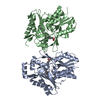
 X-RAY DIFFRACTION /
  Authors
 United States, 4items
  Citation
 Structure visualization
 Downloads & links
 Other downloads


 PDBj
PDBj Assembly


 Mass: 507.181 Da / Num. of mol.: 2 / Source method: obtained synthetically / Formula: C10H16N5O13P3 / Comment: ATP, energy-carrying molecule*YM
 Mass: 24.305 Da / Num. of mol.: 2 / Source method: obtained synthetically / Formula: Mg
 Mass: 96.063 Da / Num. of mol.: 5 / Source method: obtained synthetically / Formula: SO4
 Sample preparation
 Processing