 Movie
Movie Controller

[English] 日本語
 Yorodumi
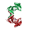
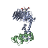
- PDB-4o4b: Crystal Structure of an Inositol hexakisphosphate kinase EhIP6KA ... -
+ Open data

- Basic information
| Entry | Database: PDB / ID: 4o4b | ||||||
|---|---|---|---|---|---|---|---|
| Title | Crystal Structure of an Inositol hexakisphosphate kinase EhIP6KA as a fusion protein with maltose binding protein | ||||||
 Components Components |
| ||||||
 Keywords Keywords | TRANSPORT PROTEIN/Transferase / PDKG kinase / TRANSPORT PROTEIN-Transferase complex | ||||||
| Function / homology |  Function and homology information Function and homology informationTransferases; Transferring phosphorus-containing groups / inositol hexakisphosphate kinase activity / inositol phosphate biosynthetic process / phosphatidylinositol phosphate biosynthetic process / ATP binding / metal ion binding / nucleus / cytoplasm Similarity search - Function | ||||||
| Biological species |    Entamoeba histolytica (eukaryote) Entamoeba histolytica (eukaryote) | ||||||
| Method |  X-RAY DIFFRACTION / SYNCHROTRON / MOLECULAR REPLACEMENT / Resolution: 1.8 Å X-RAY DIFFRACTION / SYNCHROTRON / MOLECULAR REPLACEMENT / Resolution: 1.8 Å | ||||||
 Authors Authors | Wang, H. / Shears, S.B. | ||||||
 Citation Citation | Journal: Nat Commun / Year: 2014 Title: IP6K structure and the molecular determinants of catalytic specificity in an inositol phosphate kinase family. Authors: Wang, H. / DeRose, E.F. / London, R.E. / Shears, S.B. | ||||||
| History |
|
- Structure visualization
| Structure viewer | Molecule: MolmilJmol/JSmol |
|---|
- Downloads & links
-Download
| PDBx/mmCIF format | 4o4b.cif.gz | 272.9 KB | Display | PDBx/mmCIF format |
|---|---|---|---|---|
| PDB format | pdb4o4b.ent.gz | 218.6 KB | Display | PDB format |
| PDBx/mmJSON format | 4o4b.json.gz | Tree view | PDBx/mmJSON format | |
| Others |  Other downloads Other downloads |
-Validation report
| Arichive directory | https://data.pdbj.org/pub/pdb/validation_reports/o4/4o4bftp://data.pdbj.org/pub/pdb/validation_reports/o4/4o4b | HTTPS FTP |
|---|
-Related structure data
| Related structure data | 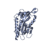 4o4cC  4o4dC 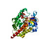 4o4eC  4o4fC 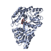 1ez9S 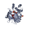 1w2cS C: citing same article ( S: Starting model for refinement |
|---|---|
| Similar structure data |


-Links
 PDBj
PDBj- Assembly
| Deposited unit | 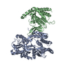
| ||||||||
|---|---|---|---|---|---|---|---|---|---|
| 1 |
| ||||||||
| Unit cell |
|
-Components
| #1: Protein | Mass: 43451.062 Da / Num. of mol.: 1 / Fragment: UNP residues 27-392 Source method: isolated from a genetically manipulated source Source: (gene. exp.) |
|---|---|
| #2: Protein | Mass: 29683.938 Da / Num. of mol.: 1 / Fragment: UNP residues 32-270 Source method: isolated from a genetically manipulated source Source: (gene. exp.) Entamoeba histolytica (eukaryote) / Strain: HM-1:IMSS / Gene: EHI7A_103520 / Plasmid: pDest566 / Production host: References: UniProt: N9UNA8, inositol-hexakisphosphate 5-kinase |
| #3: Water | ChemComp-HOH /  Mass: 18.015 Da / Num. of mol.: 654 / Source method: isolated from a natural source / Formula: H2O Mass: 18.015 Da / Num. of mol.: 654 / Source method: isolated from a natural source / Formula: H2O |
-Experimental details
-Experiment
| Experiment | Method: X-RAY DIFFRACTION / Number of used crystals: 1 |
|---|
- Sample preparation
| Crystal | Density Matthews: 2.45 Å3/Da / Density % sol: 49.84 % |
|---|---|
| Crystal grow | Temperature: 291 K / Method: vapor diffusion, hanging drop / pH: 5.5 Details: 12% (w/v) PEG 3350, 50 mM NaH2PO4, pH 5.5, VAPOR DIFFUSION, HANGING DROP, temperature 291K |
-Data collection
| Diffraction | Mean temperature: 100 K |
|---|---|
| Diffraction source | Source: SYNCHROTRON / Site: APS  / Beamline: 22-BM / Wavelength: 1 Å / Beamline: 22-BM / Wavelength: 1 Å |
| Detector | Type: MARMOSAIC 225 mm CCD / Detector: CCD / Date: Feb 5, 2013 |
| Radiation | Monochromator: SAGITALLY FOCUSED Si(111) / Protocol: SINGLE WAVELENGTH / Monochromatic (M) / Laue (L): M / Scattering type: x-ray |
| Radiation wavelength | Wavelength: 1 Å / Relative weight: 1 |
| Reflection | Resolution: 1.8→50 Å / Num. all: 67725 / Num. obs: 67725 / % possible obs: 99.4 % / Observed criterion σ(F): 3 / Observed criterion σ(I): 0 / Redundancy: 5 % / Rsym value: 0.057 / Net I/σ(I): 39.6 |
| Reflection shell | Resolution: 1.8→1.83 Å / Redundancy: 4.8 % / Mean I/σ(I) obs: 3.1 / Num. unique all: 3301 / Rsym value: 0.457 / % possible all: 99.4 |
- Processing
| Software |
| ||||||||||||||||||||||||||||||||||||||||||||||||||||||||||||||||||||||||||||||||||||||||||||||||||||||||||||||||||||||||||||||||||||||||||||||||||||||||||||||||||||||||||||||||||||||
|---|---|---|---|---|---|---|---|---|---|---|---|---|---|---|---|---|---|---|---|---|---|---|---|---|---|---|---|---|---|---|---|---|---|---|---|---|---|---|---|---|---|---|---|---|---|---|---|---|---|---|---|---|---|---|---|---|---|---|---|---|---|---|---|---|---|---|---|---|---|---|---|---|---|---|---|---|---|---|---|---|---|---|---|---|---|---|---|---|---|---|---|---|---|---|---|---|---|---|---|---|---|---|---|---|---|---|---|---|---|---|---|---|---|---|---|---|---|---|---|---|---|---|---|---|---|---|---|---|---|---|---|---|---|---|---|---|---|---|---|---|---|---|---|---|---|---|---|---|---|---|---|---|---|---|---|---|---|---|---|---|---|---|---|---|---|---|---|---|---|---|---|---|---|---|---|---|---|---|---|---|---|---|---|
| Refinement | Method to determine structure: MOLECULAR REPLACEMENT Starting model: PDB ENTRIES 1EZ9 AND 1W2C Resolution: 1.8→36.97 Å / Cor.coef. Fo:Fc: 0.958 / Cor.coef. Fo:Fc free: 0.946 / SU B: 5.902 / SU ML: 0.087 / Isotropic thermal model: Isotropic / Cross valid method: THROUGHOUT / σ(F): 0 / ESU R: 0.133 / ESU R Free: 0.12 / Stereochemistry target values: MAXIMUM LIKELIHOOD / Details: HYDROGENS HAVE BEEN ADDED IN THE RIDING POSITIONS
| ||||||||||||||||||||||||||||||||||||||||||||||||||||||||||||||||||||||||||||||||||||||||||||||||||||||||||||||||||||||||||||||||||||||||||||||||||||||||||||||||||||||||||||||||||||||
| Solvent computation | Ion probe radii: 0.8 Å / Shrinkage radii: 0.8 Å / VDW probe radii: 1.2 Å / Solvent model: MASK | ||||||||||||||||||||||||||||||||||||||||||||||||||||||||||||||||||||||||||||||||||||||||||||||||||||||||||||||||||||||||||||||||||||||||||||||||||||||||||||||||||||||||||||||||||||||
| Displacement parameters | Biso mean: 35.793 Å2
| ||||||||||||||||||||||||||||||||||||||||||||||||||||||||||||||||||||||||||||||||||||||||||||||||||||||||||||||||||||||||||||||||||||||||||||||||||||||||||||||||||||||||||||||||||||||
| Refinement step | Cycle: LAST / Resolution: 1.8→36.97 Å
| ||||||||||||||||||||||||||||||||||||||||||||||||||||||||||||||||||||||||||||||||||||||||||||||||||||||||||||||||||||||||||||||||||||||||||||||||||||||||||||||||||||||||||||||||||||||
| Refine LS restraints |
|