 Movie
Movie Controller


+ Open data

- Basic information
| Entry | Database: PDB / ID: 6qm2 | ||||||
|---|---|---|---|---|---|---|---|
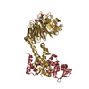
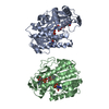
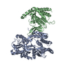
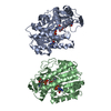
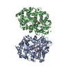
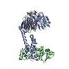
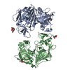
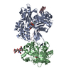
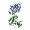
| Title | NlaIV restriction endonuclease | ||||||
 Components Components | Type-2 restriction enzyme NlaIV | ||||||
 Keywords Keywords | HYDROLASE / RESTRICTION ENDONUCLEASE / NLAIV / BLUNT END CUTTER | ||||||
| Function / homology | Type-2 restriction enzyme NlaIV / NgoBV restriction endonuclease / type II site-specific deoxyribonuclease / type II site-specific deoxyribonuclease activity / DNA restriction-modification system / : / Type II restriction enzyme NlaIV Function and homology information Function and homology information | ||||||
| Biological species |  Neisseria lactamica (bacteria) Neisseria lactamica (bacteria) | ||||||
| Method |  X-RAY DIFFRACTION / SYNCHROTRON / MAD / Resolution: 2.8 Å X-RAY DIFFRACTION / SYNCHROTRON / MAD / Resolution: 2.8 Å | ||||||
 Authors Authors | Czapinska, H. / Siwek, W. / Szczepanowski, R.H. / Bujnicki, J.M. / Bochtler, M. / Skowronek, K. | ||||||
 Citation Citation | Journal: J.Mol.Biol. / Year: 2019 Title: Crystal Structure and Directed Evolution of Specificity of NlaIV Restriction Endonuclease. Authors: Czapinska, H. / Siwek, W. / Szczepanowski, R.H. / Bujnicki, J.M. / Bochtler, M. / Skowronek, K.J. #1: Journal: Protein Eng. Des. Sel. / Year: 2005 Title: A theoretical model of restriction endonuclease NlaIV in complex with DNA, predicted by fold recognition and validated by site-directed mutagenesis and circular dichroism spectroscopy. Authors: Chmiel, A.A. / Radlinska, M. / Pawlak, S.D. / Krowarsch, D. / Bujnicki, J.M. / Skowronek, K.J. | ||||||
| History |
|
- Structure visualization
| Structure viewer | Molecule: MolmilJmol/JSmol |
|---|
- Downloads & links
-Download
| PDBx/mmCIF format | 6qm2.cif.gz | 69.7 KB | Display | PDBx/mmCIF format |
|---|---|---|---|---|
| PDB format | pdb6qm2.ent.gz | 50.5 KB | Display | PDB format |
| PDBx/mmJSON format | 6qm2.json.gz | Tree view | PDBx/mmJSON format | |
| Others |  Other downloads Other downloads |
-Validation report
| Arichive directory | https://data.pdbj.org/pub/pdb/validation_reports/qm/6qm2ftp://data.pdbj.org/pub/pdb/validation_reports/qm/6qm2 | HTTPS FTP |
|---|
-Related structure data
| Similar structure data |
|---|

-Links
 PDBj
PDBj

- Assembly
| Deposited unit | 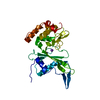
| ||||||||
|---|---|---|---|---|---|---|---|---|---|
| 1 | 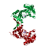
| ||||||||
| Unit cell |
|
-Components
| #1: Protein | Mass: 29153.611 Da / Num. of mol.: 1 Source method: isolated from a genetically manipulated source Source: (gene. exp.) Neisseria lactamica (bacteria) / Strain: NRCC 2118 / Gene: nlaIVR / Plasmid: PET28a / Production host: References: UniProt: P50183, type II site-specific deoxyribonuclease |
|---|---|
| #2: Chemical | ChemComp-NA /   Mass: 22.990 Da / Num. of mol.: 1 / Source method: obtained synthetically / Formula: Na Mass: 22.990 Da / Num. of mol.: 1 / Source method: obtained synthetically / Formula: Na |
| #3: Chemical | ChemComp-K /   Mass: 39.098 Da / Num. of mol.: 1 / Source method: obtained synthetically / Formula: K Mass: 39.098 Da / Num. of mol.: 1 / Source method: obtained synthetically / Formula: K |
| #4: Water | ChemComp-HOH /  Mass: 18.015 Da / Num. of mol.: 123 / Source method: isolated from a natural source / Formula: H2O Mass: 18.015 Da / Num. of mol.: 123 / Source method: isolated from a natural source / Formula: H2O |
-Experimental details
-Experiment
| Experiment | Method: X-RAY DIFFRACTION / Number of used crystals: 1 |
|---|
- Sample preparation
| Crystal grow | Temperature: 294 K / Method: vapor diffusion, sitting drop / pH: 5 Details: 10 mg/ml of the enzyme in 15 % glycerol, 50 mM NaOH/HEPES pH 7,5, 50 mM KCl, 10 mM DTT and 5 mM CaCl2 was mixed in 1:1 ratio with double stranded DNA composed of the 5'-ATGGTACCTGC-3' and 5'- ...Details: 10 mg/ml of the enzyme in 15 % glycerol, 50 mM NaOH/HEPES pH 7,5, 50 mM KCl, 10 mM DTT and 5 mM CaCl2 was mixed in 1:1 ratio with double stranded DNA composed of the 5'-ATGGTACCTGC-3' and 5'-CAGGTACCATG-3' strands. The protein-DNA solutions were in turn mixed in 1:1 ratio with the precipitant solution containing 2 M NaCl and 100 mM citric acid, pH 5. PH range: 5 |
|---|
-Data collection
| Diffraction | Mean temperature: 100 K / Serial crystal experiment: N |
|---|---|
| Diffraction source | Source: SYNCHROTRON / Site: MPG/DESY, HAMBURG  / Beamline: BW6 / Wavelength: 1.05 Å / Beamline: BW6 / Wavelength: 1.05 Å |
| Detector | Type: MARRESEARCH / Detector: CCD / Date: Dec 17, 2008 / Details: BENT MIRROR |
| Radiation | Monochromator: TRIANGULAR MONOCHROMATOR / Protocol: SINGLE WAVELENGTH / Monochromatic (M) / Laue (L): M / Scattering type: x-ray |
| Radiation wavelength | Wavelength: 1.05 Å / Relative weight: 1 |
| Reflection | Resolution: 2.8→30 Å / Num. obs: 18997 / % possible obs: 98.5 % / Redundancy: 7.5 % / Rmerge(I) obs: 0.043 / Rsym value: 0.04 / Net I/σ(I): 28.92 |
| Reflection shell | Resolution: 2.8→2.97 Å / Redundancy: 7.7 % / Rmerge(I) obs: 1.052 / Mean I/σ(I) obs: 1.72 / Rsym value: 0.986 / % possible all: 98.6 |
- Processing
| Software |
| ||||||||||||||||||||||||||||||||||||||||||||||||||||||||||||||||||||||||||||||||||||||||||||||||||||||||||||||||||||||||||||||||||||||||||||||||||||||||||||||||||||||||||||||||||||||
|---|---|---|---|---|---|---|---|---|---|---|---|---|---|---|---|---|---|---|---|---|---|---|---|---|---|---|---|---|---|---|---|---|---|---|---|---|---|---|---|---|---|---|---|---|---|---|---|---|---|---|---|---|---|---|---|---|---|---|---|---|---|---|---|---|---|---|---|---|---|---|---|---|---|---|---|---|---|---|---|---|---|---|---|---|---|---|---|---|---|---|---|---|---|---|---|---|---|---|---|---|---|---|---|---|---|---|---|---|---|---|---|---|---|---|---|---|---|---|---|---|---|---|---|---|---|---|---|---|---|---|---|---|---|---|---|---|---|---|---|---|---|---|---|---|---|---|---|---|---|---|---|---|---|---|---|---|---|---|---|---|---|---|---|---|---|---|---|---|---|---|---|---|---|---|---|---|---|---|---|---|---|---|---|
| Refinement | Method to determine structure: MAD / Resolution: 2.8→29.72 Å / Cor.coef. Fo:Fc: 0.966 / Cor.coef. Fo:Fc free: 0.956 / SU B: 11.197 / SU ML: 0.189 / Cross valid method: THROUGHOUT / ESU R: 0.267 / ESU R Free: 0.221 Details: HYDROGENS HAVE BEEN ADDED IN THE RIDING POSITIONS. The ION IDENTITY HAS BEEN ASSIGNED TENTATIVELY.
| ||||||||||||||||||||||||||||||||||||||||||||||||||||||||||||||||||||||||||||||||||||||||||||||||||||||||||||||||||||||||||||||||||||||||||||||||||||||||||||||||||||||||||||||||||||||
| Solvent computation | Ion probe radii: 0.8 Å / Shrinkage radii: 0.8 Å / VDW probe radii: 1.2 Å | ||||||||||||||||||||||||||||||||||||||||||||||||||||||||||||||||||||||||||||||||||||||||||||||||||||||||||||||||||||||||||||||||||||||||||||||||||||||||||||||||||||||||||||||||||||||
| Displacement parameters | Biso mean: 115.889 Å2
| ||||||||||||||||||||||||||||||||||||||||||||||||||||||||||||||||||||||||||||||||||||||||||||||||||||||||||||||||||||||||||||||||||||||||||||||||||||||||||||||||||||||||||||||||||||||
| Refinement step | Cycle: LAST / Resolution: 2.8→29.72 Å
| ||||||||||||||||||||||||||||||||||||||||||||||||||||||||||||||||||||||||||||||||||||||||||||||||||||||||||||||||||||||||||||||||||||||||||||||||||||||||||||||||||||||||||||||||||||||
| Refine LS restraints |
|