 Movie
Movie Controller

[English] 日本語
 Yorodumi
- PDB-6mh9: The crystal structure of the Staphylococcus aureus Fatty acid Kin... -
+ Open data

- Basic information
| Entry | Database: PDB / ID: 6mh9 | ||||||
|---|---|---|---|---|---|---|---|
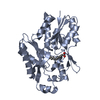
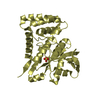
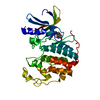
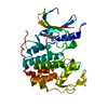
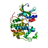
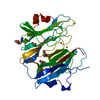
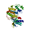
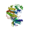
| Title | The crystal structure of the Staphylococcus aureus Fatty acid Kinase (Fak) B1 protein A121I mutant to 2.02 Angstrom resolution | ||||||
 Components Components | Fatty Acid Kinase (Fak) B1 protein | ||||||
 Keywords Keywords | TRANSFERASE / Staphylococcus aureus / FakB1 / mutant TRANSFERASE | ||||||
| Function / homology |  Function and homology information Function and homology information | ||||||
| Biological species |   Staphylococcus aureus (bacteria) Staphylococcus aureus (bacteria) | ||||||
| Method |  X-RAY DIFFRACTION / SYNCHROTRON / MOLECULAR REPLACEMENT / Resolution: 2.02 Å X-RAY DIFFRACTION / SYNCHROTRON / MOLECULAR REPLACEMENT / Resolution: 2.02 Å | ||||||
 Authors Authors | Cuypers, M.G. / Ericson, M. / Subramanian, C. / White, S.W. / Rock, C.O. | ||||||
 Citation Citation | Journal: J.Biol.Chem. / Year: 2022 Title: Identification of structural transitions in bacterial fatty acid binding proteins that permit ligand entry and exit at membranes. Authors: Gullett, J.M. / Cuypers, M.G. / Grace, C.R. / Pant, S. / Subramanian, C. / Tajkhorshid, E. / Rock, C.O. / White, S.W. | ||||||
| History |
|
- Structure visualization
| Structure viewer | Molecule: MolmilJmol/JSmol |
|---|
- Downloads & links
-Download
| PDBx/mmCIF format | 6mh9.cif.gz | 130.9 KB | Display | PDBx/mmCIF format |
|---|---|---|---|---|
| PDB format | pdb6mh9.ent.gz | 100.2 KB | Display | PDB format |
| PDBx/mmJSON format | 6mh9.json.gz | Tree view | PDBx/mmJSON format | |
| Others |  Other downloads Other downloads |
-Validation report
| Arichive directory | https://data.pdbj.org/pub/pdb/validation_reports/mh/6mh9ftp://data.pdbj.org/pub/pdb/validation_reports/mh/6mh9 | HTTPS FTP |
|---|
-Related structure data
| Related structure data |  6nm1C  7sclC  7sg3C 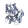 5utoS S: Starting model for refinement C: citing same article ( |
|---|---|
| Similar structure data |


-Links
 PDBj
PDBj


- Assembly
| Deposited unit | 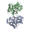
| ||||||||
|---|---|---|---|---|---|---|---|---|---|
| 1 | 
| ||||||||
| 2 | 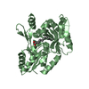
| ||||||||
| Unit cell |
|
-Components
| #1: Protein | Mass: 32281.729 Da / Num. of mol.: 2 / Mutation: A121I Source method: isolated from a genetically manipulated source Source: (gene. exp.) Staphylococcus aureus (bacteria)Gene: BTN44_08795, CV021_09110, EP54_02745, EQ90_03735, ERS072840_01626, HMPREF3211_01094 Production host: #2: Chemical |   Mass: 256.424 Da / Num. of mol.: 2 / Source method: obtained synthetically / Formula: C16H32O2 / Feature type: SUBJECT OF INVESTIGATION Mass: 256.424 Da / Num. of mol.: 2 / Source method: obtained synthetically / Formula: C16H32O2 / Feature type: SUBJECT OF INVESTIGATION#3: Water | ChemComp-HOH / |  Mass: 18.015 Da / Num. of mol.: 207 / Source method: isolated from a natural source / Formula: H2O Mass: 18.015 Da / Num. of mol.: 207 / Source method: isolated from a natural source / Formula: H2O |
|---|
-Experimental details
-Experiment
| Experiment | Method: X-RAY DIFFRACTION / Number of used crystals: 1 |
|---|
- Sample preparation
| Crystal | Density Matthews: 2.11 Å3/Da / Density % sol: 41.68 % |
|---|---|
| Crystal grow | Temperature: 293 K / Method: vapor diffusion, hanging drop Details: PH 6.5 0.1M MES/IMIDAZOLE, 12.5% PEG1000, 12.5% PEG3350, 12.5% MPD, 0.03M NANO3, 0.03M NA2HPO4, 0.03M (NH4)2 SO4 Temp details: stable room temperature |
-Data collection
| Diffraction | Mean temperature: 100 K |
|---|---|
| Diffraction source | Source: SYNCHROTRON / Site: APS  / Beamline: 22-ID / Wavelength: 1.6039 Å / Beamline: 22-ID / Wavelength: 1.6039 Å |
| Detector | Type: RAYONIX MX300-HS / Detector: CCD / Date: Feb 26, 2017 |
| Radiation | Protocol: SINGLE WAVELENGTH / Monochromatic (M) / Laue (L): M / Scattering type: x-ray |
| Radiation wavelength | Wavelength: 1.6039 Å / Relative weight: 1 |
| Reflection | Resolution: 2.02→83.29 Å / Num. obs: 34458 / % possible obs: 94.4 % / Redundancy: 3.8 % / Biso Wilson estimate: 34.8 Å2 / CC1/2: 0.997 / Rmerge(I) obs: 0.066 / Rpim(I) all: 0.039 / Rrim(I) all: 0.077 / Net I/σ(I): 12.3 |
| Reflection shell | Resolution: 2.02→2.07 Å / Redundancy: 3.7 % / Rmerge(I) obs: 0.585 / Num. unique obs: 2509 / CC1/2: 0.724 / Rpim(I) all: 0.438 / Rrim(I) all: 0.849 / % possible all: 91.3 |
- Processing
| Software |
| |||||||||||||||||||||||||||||||||||||||||||||||||||||||||||||||||||||||||||||||||||||||||||
|---|---|---|---|---|---|---|---|---|---|---|---|---|---|---|---|---|---|---|---|---|---|---|---|---|---|---|---|---|---|---|---|---|---|---|---|---|---|---|---|---|---|---|---|---|---|---|---|---|---|---|---|---|---|---|---|---|---|---|---|---|---|---|---|---|---|---|---|---|---|---|---|---|---|---|---|---|---|---|---|---|---|---|---|---|---|---|---|---|---|---|---|---|
| Refinement | Method to determine structure: MOLECULAR REPLACEMENT Starting model: 5UTO Resolution: 2.02→48.441 Å / SU ML: 0.27 / Cross valid method: FREE R-VALUE / σ(F): 1.98 / Phase error: 30.09
| |||||||||||||||||||||||||||||||||||||||||||||||||||||||||||||||||||||||||||||||||||||||||||
| Solvent computation | Shrinkage radii: 0.9 Å / VDW probe radii: 1.11 Å | |||||||||||||||||||||||||||||||||||||||||||||||||||||||||||||||||||||||||||||||||||||||||||
| Refinement step | Cycle: LAST / Resolution: 2.02→48.441 Å
| |||||||||||||||||||||||||||||||||||||||||||||||||||||||||||||||||||||||||||||||||||||||||||
| Refine LS restraints |
| |||||||||||||||||||||||||||||||||||||||||||||||||||||||||||||||||||||||||||||||||||||||||||
| LS refinement shell |
|
