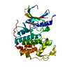
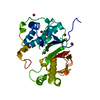
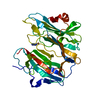
peptidylglycine monooxygenase / peptidylamidoglycolate lyase / peptide amidation / peptidylglycine monooxygenase activity / peptidylamidoglycolate lyase activity / fatty acid primary amide biosynthetic process / ovulation cycle process / toxin metabolic process / long-chain fatty acid metabolic process / peptide metabolic process ...peptidylglycine monooxygenase / peptidylamidoglycolate lyase / peptide amidation / peptidylglycine monooxygenase activity / peptidylamidoglycolate lyase activity / fatty acid primary amide biosynthetic process / ovulation cycle process / toxin metabolic process / long-chain fatty acid metabolic process / peptide metabolic process / mitotic chromosome condensation / response to pH / L-ascorbic acid binding / response to zinc ion / response to copper ion / transport vesicle membrane / limb development / maternal process involved in female pregnancy / condensed chromosome / lactation / secretory granule / response to glucocorticoid / regulation of actin cytoskeleton organization / trans-Golgi network / response to estradiol / heart development / cellular response to oxidative stress / perikaryon / proteasome-mediated ubiquitin-dependent protein catabolic process / response to hypoxia / response to xenobiotic stimulus / copper ion binding / neuronal cell body / calcium ion binding / chromatin binding / regulation of transcription by RNA polymerase II / protein kinase binding / chromatin / perinuclear region of cytoplasm / cell surface / : / extracellular region / zinc ion binding / identical protein binding Similarity search - Function
Copper type II, ascorbate-dependent monooxygenase, N-terminal domain / Peptidylglycine alpha-hydroxylating monooxygenase/peptidyl-hydroxyglycine alpha-amidating lyase / Jelly Rolls - #230 / Copper type II, ascorbate-dependent monooxygenase, histidine-cluster-2 conserved site / Copper type II, ascorbate-dependent monooxygenases signature 2. / Copper type II, ascorbate-dependent monooxygenase, N-terminal / Copper type II, ascorbate-dependent monooxygenase, histidine-cluster-1 conserved site / Copper type II ascorbate-dependent monooxygenase, C-terminal / Copper type II, ascorbate-dependent monooxygenase, N-terminal domain superfamily / Copper type II ascorbate-dependent monooxygenase, N-terminal domain ...Copper type II, ascorbate-dependent monooxygenase, N-terminal domain / Peptidylglycine alpha-hydroxylating monooxygenase/peptidyl-hydroxyglycine alpha-amidating lyase / Jelly Rolls - #230 / Copper type II, ascorbate-dependent monooxygenase, histidine-cluster-2 conserved site / Copper type II, ascorbate-dependent monooxygenases signature 2. / Copper type II, ascorbate-dependent monooxygenase, N-terminal / Copper type II, ascorbate-dependent monooxygenase, histidine-cluster-1 conserved site / Copper type II ascorbate-dependent monooxygenase, C-terminal / Copper type II, ascorbate-dependent monooxygenase, N-terminal domain superfamily / Copper type II ascorbate-dependent monooxygenase, N-terminal domain / Copper type II ascorbate-dependent monooxygenase, C-terminal domain / Copper type II, ascorbate-dependent monooxygenases signature 1. / PHM/PNGase F domain superfamily / Copper type II, ascorbate-dependent monooxygenase-like, C-terminal / NHL repeat profile. / NHL repeat / NHL repeat / Six-bladed beta-propeller, TolB-like / Jelly Rolls / Sandwich / Mainly Beta Similarity search - Domain/homology
In the structure databanks used in Yorodumi, some data are registered as the other names, "COVID-19 virus" and "2019-nCoV". Here are the details of the virus and the list of structure data.
Jan 31, 2019. EMDB accession codes are about to change! (news from PDBe EMDB page)
EMDB accession codes are about to change! (news from PDBe EMDB page)
The allocation of 4 digits for EMDB accession codes will soon come to an end. Whilst these codes will remain in use, new EMDB accession codes will include an additional digit and will expand incrementally as the available range of codes is exhausted. The current 4-digit format prefixed with “EMD-” (i.e. EMD-XXXX) will advance to a 5-digit format (i.e. EMD-XXXXX), and so on. It is currently estimated that the 4-digit codes will be depleted around Spring 2019, at which point the 5-digit format will come into force.
The EM Navigator/Yorodumi systems omit the EMD- prefix.
Related info.:Q: What is EMD? / ID/Accession-code notation in Yorodumi/EM Navigator
Yorodumi is a browser for structure data from EMDB, PDB, SASBDB, etc.
This page is also the successor to EM Navigator detail page, and also detail information page/front-end page for Omokage search.
The word "yorodu" (or yorozu) is an old Japanese word meaning "ten thousand". "mi" (miru) is to see.
Related info.:EMDB / PDB / SASBDB / Comparison of 3 databanks / Yorodumi Search / Aug 31, 2016. New EM Navigator & Yorodumi / Yorodumi Papers / Jmol/JSmol / Function and homology information / Changes in new EM Navigator and Yorodumi
 Movie
Movie Controller


 Open data
 Basic information
 Components
 Keywords
 Function and homology information
 X-RAY DIFFRACTION /
  Authors
 Citation
 Structure visualization
 Downloads & links
 Other downloads
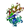


 PDBj
PDBj


 Assembly
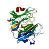
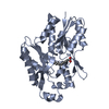
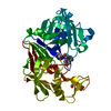
 Cricetulus griseus (Chinese hamster) / References: UniProt: P14925, peptidylglycine monooxygenase
 Mass: 63.546 Da / Num. of mol.: 3 / Source method: obtained synthetically / Formula: Cu
 Mass: 42.020 Da / Num. of mol.: 1 / Source method: obtained synthetically / Formula: N3
 Mass: 92.094 Da / Num. of mol.: 4 / Source method: obtained synthetically / Formula: C3H8O3
 Mass: 18.015 Da / Num. of mol.: 226 / Source method: isolated from a natural source / Formula: H2O
 Sample preparation
 / Beamline: X4A / Wavelength: 0.9793
  Processing