 Movie
Movie Controller


+ Open data

- Basic information
| Entry | Database: PDB / ID: 1mn4 | ||||||
|---|---|---|---|---|---|---|---|
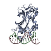
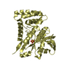
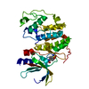
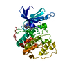
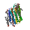
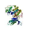
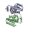
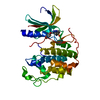
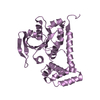
| Title | Structure of Ndt80 (Residues 59-340) DNA-binding domain core | ||||||
 Components Components | NDT80 PROTEIN | ||||||
 Keywords Keywords | TRANSCRIPTION / IG FOLD / PROTEIN-DNA COMPLEX | ||||||
| Function / homology |  Function and homology information Function and homology informationsporulation / nuclear chromosome / meiotic cell cycle / sequence-specific DNA binding / DNA-binding transcription factor activity / cell division / positive regulation of transcription by RNA polymerase II / DNA-templated transcription Similarity search - Function | ||||||
| Biological species |  | ||||||
| Method |  X-RAY DIFFRACTION / SYNCHROTRON / MAD / Resolution: 2.2 Å X-RAY DIFFRACTION / SYNCHROTRON / MAD / Resolution: 2.2 Å | ||||||
 Authors Authors | Lamoureux, J.S. / Stuart, D. / Tsang, R. / Wu, C. / Glover, J.N.M. | ||||||
 Citation Citation | Journal: Embo J. / Year: 2002 Title: Structure of the sporulation-specific transcription factor Ndt80 bound to DNA Authors: Lamoureux, J.S. / Stuart, D. / Tsang, R. / Wu, C. / Glover, J.N.M. | ||||||
| History |
|
- Structure visualization
| Structure viewer | Molecule: MolmilJmol/JSmol |
|---|
- Downloads & links
-Download
| PDBx/mmCIF format | 1mn4.cif.gz | 64.1 KB | Display | PDBx/mmCIF format |
|---|---|---|---|---|
| PDB format | pdb1mn4.ent.gz | 46.6 KB | Display | PDB format |
| PDBx/mmJSON format | 1mn4.json.gz | Tree view | PDBx/mmJSON format | |
| Others |  Other downloads Other downloads |
-Validation report
| Arichive directory | https://data.pdbj.org/pub/pdb/validation_reports/mn/1mn4ftp://data.pdbj.org/pub/pdb/validation_reports/mn/1mn4 | HTTPS FTP |
|---|
-Related structure data


-Links
 PDBj
PDBj


- Assembly
| Deposited unit | 
| ||||||||
|---|---|---|---|---|---|---|---|---|---|
| 1 |
| ||||||||
| Unit cell |
|
-Components
| #1: Protein | Mass: 32385.701 Da / Num. of mol.: 1 / Fragment: DNA-binding core (Residues 59-340) Source method: isolated from a genetically manipulated source Source: (gene. exp.) Gene: NDT80 / Plasmid: pGEX-6P1 / Species (production host): Escherichia coli / Production host:  |
|---|---|
| #2: Water | ChemComp-HOH /  Mass: 18.015 Da / Num. of mol.: 89 / Source method: isolated from a natural source / Formula: H2O Mass: 18.015 Da / Num. of mol.: 89 / Source method: isolated from a natural source / Formula: H2O |
-Experimental details
-Experiment
| Experiment | Method: X-RAY DIFFRACTION / Number of used crystals: 1 |
|---|
- Sample preparation
| Crystal | Density Matthews: 1.91 Å3/Da / Density % sol: 35.58 % | |||||||||||||||||||||||||||||||||||||||||||||||||
|---|---|---|---|---|---|---|---|---|---|---|---|---|---|---|---|---|---|---|---|---|---|---|---|---|---|---|---|---|---|---|---|---|---|---|---|---|---|---|---|---|---|---|---|---|---|---|---|---|---|---|
| Crystal grow | Temperature: 293 K / Method: vapor diffusion, hanging drop / pH: 7 Details: PEG 4000, sodium acetate, magnesium chloride, spermine, 20mer DNA, pH 7.0, VAPOR DIFFUSION, HANGING DROP, temperature 293K | |||||||||||||||||||||||||||||||||||||||||||||||||
| Crystal grow | *PLUS Temperature: 20 ℃ / pH: 5.5 | |||||||||||||||||||||||||||||||||||||||||||||||||
| Components of the solutions | *PLUS
|
-Data collection
| Diffraction | Mean temperature: 105 K |
|---|---|
| Diffraction source | Source: SYNCHROTRON / Site: APS  / Beamline: 14-BM-D / Wavelength: 0.9563 Å / Beamline: 14-BM-D / Wavelength: 0.9563 Å |
| Detector | Type: ADSC QUANTUM 4 / Detector: CCD / Date: May 21, 2002 / Details: bent cylindrical Si-mirror (Rh coating) |
| Radiation | Monochromator: Si(111) double-crystal / Protocol: MAD / Monochromatic (M) / Laue (L): M / Scattering type: x-ray |
| Radiation wavelength | Wavelength: 0.9563 Å / Relative weight: 1 |
| Reflection | Resolution: 2.2→14.95 Å / Num. all: 12530 / Num. obs: 13292 / % possible obs: 98.1 % / Observed criterion σ(F): 0 / Observed criterion σ(I): -3 / Redundancy: 11.7 % / Biso Wilson estimate: 14.3 Å2 / Rsym value: 0.076 / Net I/σ(I): 20.9 |
| Reflection shell | Resolution: 2.2→2.25 Å / Redundancy: 5.4 % / Mean I/σ(I) obs: 7.9 / Rsym value: 0.221 / % possible all: 100 |
| Reflection | *PLUS Lowest resolution: 40 Å / Num. obs: 12292 / Num. measured all: 144249 / Rmerge(I) obs: 0.076 |
| Reflection shell | *PLUS % possible obs: 100 % / Rmerge(I) obs: 0.221 |
- Processing
| Software |
| ||||||||||||||||||||||||||||
|---|---|---|---|---|---|---|---|---|---|---|---|---|---|---|---|---|---|---|---|---|---|---|---|---|---|---|---|---|---|
| Refinement | Method to determine structure: MAD / Resolution: 2.2→14.95 Å / Isotropic thermal model: anisotropic / Cross valid method: THROUGHOUT / σ(F): 0 / σ(I): 0 / Stereochemistry target values: Engh & Huber
| ||||||||||||||||||||||||||||
| Displacement parameters |
| ||||||||||||||||||||||||||||
| Refine analyze | Luzzati coordinate error obs: 0.27 Å / Luzzati d res low obs: 5 Å / Luzzati sigma a obs: 0.21 Å | ||||||||||||||||||||||||||||
| Refinement step | Cycle: LAST / Resolution: 2.2→14.95 Å
| ||||||||||||||||||||||||||||
| Refine LS restraints |
| ||||||||||||||||||||||||||||
| Xplor file |
| ||||||||||||||||||||||||||||
| Refinement | *PLUS Lowest resolution: 40 Å / Rfactor all: 0.229 / Rfactor Rfree: 0.268 / Rfactor Rwork: 0.226 / % reflection Rfree: 5 % | ||||||||||||||||||||||||||||
| Solvent computation | *PLUS | ||||||||||||||||||||||||||||
| Displacement parameters | *PLUS | ||||||||||||||||||||||||||||
| Refine LS restraints | *PLUS Type: c_angle_deg / Dev ideal: 1.28 |
