 ムービー
ムービー コントローラー


+ データを開く

- 基本情報
基本情報
| 登録情報 | データベース: PDB / ID: 1ucq | |||||||||
|---|---|---|---|---|---|---|---|---|---|---|
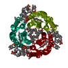
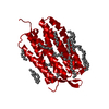
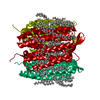
| タイトル | Crystal structure of the L intermediate of bacteriorhodopsin | |||||||||
 要素 要素 | bacteriorhodopsin | |||||||||
 キーワード キーワード | PROTON TRANSPORT / proton pump / retinal protein / membrane protein / protein-lipid complex / reaction intermediate | |||||||||
| 機能・相同性 |  機能・相同性情報 機能・相同性情報light-driven active monoatomic ion transmembrane transporter activity / photoreceptor activity / phototransduction / monoatomic ion channel activity / proton transmembrane transport / plasma membrane 類似検索 - 分子機能 | |||||||||
| 生物種 |  Halobacterium salinarum (好塩性) Halobacterium salinarum (好塩性) | |||||||||
| 手法 |  X線回折 / シンクロトロン / 分子置換 / 解像度: 2.4 Å X線回折 / シンクロトロン / 分子置換 / 解像度: 2.4 Å | |||||||||
 データ登録者 データ登録者 | Kouyama, T. / Nishikawa, T. / Tokuhisa, T. / Okumura, H. | |||||||||
 引用 引用 | ジャーナル: J.Mol.Biol. / 年: 2004 タイトル: Crystal Structure of the L Intermediate of Bacteriorhodopsin: Evidence for Vertical Translocation of a Water Molecule during the Proton Pumping Cycle. 著者: Kouyama, T. / Nishikawa, T. / Tokuhisa, T. / Okumura, H. | |||||||||
| 履歴 |
|
- 構造の表示
構造の表示
| 構造ビューア | 分子: MolmilJmol/JSmol |
|---|
- ダウンロードとリンク
-ダウンロード
| PDBx/mmCIF形式 | 1ucq.cif.gz | 68.4 KB | 表示 | PDBx/mmCIF形式 |
|---|---|---|---|---|
| PDB形式 | pdb1ucq.ent.gz | 48.8 KB | 表示 | PDB形式 |
| PDBx/mmJSON形式 | 1ucq.json.gz | ツリー表示 | PDBx/mmJSON形式 | |
| その他 |  その他のダウンロード その他のダウンロード |
-検証レポート
| アーカイブディレクトリ | https://data.pdbj.org/pub/pdb/validation_reports/uc/1ucqftp://data.pdbj.org/pub/pdb/validation_reports/uc/1ucq | HTTPS FTP |
|---|
-関連構造データ
| 関連構造データ | 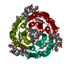 1iw6S S: 精密化の開始モデル |
|---|---|
| 類似構造データ |


-リンク
 PDBj
PDBj


- 集合体
集合体
| 登録構造単位 | 
| ||||||||
|---|---|---|---|---|---|---|---|---|---|
| 1 | 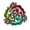
| ||||||||
| 単位格子 |
| ||||||||
| 詳細 | The biological assembly is a trimer generated from the monomer in the asymmetric unit by the operations: -y, x-y, z and -x+y, -x, z. |
-要素
-タンパク質 / 糖 , 2種, 2分子 A
| #1: タンパク質 | 分子量: 26929.500 Da / 分子数: 1 / 由来タイプ: 天然 / 由来: (天然) Halobacterium salinarum (好塩性) / 株: jw3 / 参照: UniProt: P02945 |
|---|---|
| #2: 多糖 | beta-D-galactopyranose-(1-6)-alpha-D-mannopyranose-(1-2)-alpha-D-glucopyranose |
-非ポリマー , 4種, 47分子 

| #3: 化合物 | ChemComp-RET /  分子量: 284.436 Da / 分子数: 1 / 由来タイプ: 合成 / 式: C20H28O 分子量: 284.436 Da / 分子数: 1 / 由来タイプ: 合成 / 式: C20H28O | ||||
|---|---|---|---|---|---|
| #4: 化合物 | ChemComp-L3P /  分子量: 885.179 Da / 分子数: 4 / 由来タイプ: 合成 / 式: C46H94O11P2 分子量: 885.179 Da / 分子数: 4 / 由来タイプ: 合成 / 式: C46H94O11P2#5: 化合物 | ChemComp-L2P / |  分子量: 653.157 Da / 分子数: 1 / 由来タイプ: 合成 / 式: C43H88O3 分子量: 653.157 Da / 分子数: 1 / 由来タイプ: 合成 / 式: C43H88O3#6: 水 | ChemComp-HOH / | 分子量: 18.015 Da / 分子数: 41 / 由来タイプ: 天然 / 式: H2O |
-詳細
| Has protein modification | Y |
|---|
-実験情報
-実験
| 実験 | 手法: X線回折 / 使用した結晶の数: 4 |
|---|
- 試料調製
試料調製
| 結晶 | マシュー密度: 2.89 Å3/Da / 溶媒含有率: 57.13 % | ||||||||||||||||||||||||||||||||||||||||||||||||||||||||
|---|---|---|---|---|---|---|---|---|---|---|---|---|---|---|---|---|---|---|---|---|---|---|---|---|---|---|---|---|---|---|---|---|---|---|---|---|---|---|---|---|---|---|---|---|---|---|---|---|---|---|---|---|---|---|---|---|---|
| 結晶化 | 温度: 278 K / 手法: 蒸気拡散法, シッティングドロップ法 / pH: 5.2 詳細: ammonium sulfate, pH 5.2, VAPOR DIFFUSION, SITTING DROP, temperature 278.0K | ||||||||||||||||||||||||||||||||||||||||||||||||||||||||
| 結晶化 | *PLUS 温度: 5 ℃ / 手法: 蒸気拡散法, シッティングドロップ法 | ||||||||||||||||||||||||||||||||||||||||||||||||||||||||
| 溶液の組成 | *PLUS
|
-データ収集
| 回折 | 平均測定温度: 100 K |
|---|---|
| 放射光源 | 由来: シンクロトロン / サイト: SPring-8  / ビームライン: BL40B2 / 波長: 1 Å / ビームライン: BL40B2 / 波長: 1 Å |
| 検出器 | タイプ: ADSC QUANTUM 4 / 検出器: CCD / 日付: 2003年2月16日 |
| 放射 | モノクロメーター: Si 111 / プロトコル: SINGLE WAVELENGTH / 単色(M)・ラウエ(L): M / 散乱光タイプ: x-ray |
| 放射波長 | 波長: 1 Å / 相対比: 1 |
| 反射 | 解像度: 2.4→50 Å / Num. all: 13880 / Num. obs: 13880 / % possible obs: 99.8 % / Observed criterion σ(F): 0 / Observed criterion σ(I): 0 / 冗長度: 5.7 % / Biso Wilson estimate: 50.406 Å2 / Rsym value: 0.066 / Net I/σ(I): 5.3 |
| 反射 シェル | 解像度: 2.4→2.53 Å / 冗長度: 5.8 % / Mean I/σ(I) obs: 1.5 / Num. unique all: 1986 / Rsym value: 0.492 / % possible all: 99.8 |
| 反射 | *PLUS 最低解像度: 30 Å / Rmerge(I) obs: 0.066 |
| 反射 シェル | *PLUS % possible obs: 49.2 % / Num. measured obs: 99.8 |
- 解析
解析
| ソフトウェア |
| |||||||||||||||||||||
|---|---|---|---|---|---|---|---|---|---|---|---|---|---|---|---|---|---|---|---|---|---|---|
| 精密化 | 構造決定の手法: 分子置換 開始モデル: 1iw6 解像度: 2.4→50 Å / Isotropic thermal model: Isotropic / 交差検証法: THROUGHOUT / σ(F): 0 / 立体化学のターゲット値: Engh & Huber 詳細: Diffraction data were collected for a 4:1 mixture of the ground state and the L intermediate. For refinement of the L-state structure, the contribution of the ground state was subtracted ...詳細: Diffraction data were collected for a 4:1 mixture of the ground state and the L intermediate. For refinement of the L-state structure, the contribution of the ground state was subtracted using diffraction data collected for the pure ground state.
| |||||||||||||||||||||
| 原子変位パラメータ | Biso mean: 66.4738 Å2
| |||||||||||||||||||||
| Refine analyze |
| |||||||||||||||||||||
| 精密化ステップ | サイクル: LAST / 解像度: 2.4→50 Å
| |||||||||||||||||||||
| 拘束条件 |
| |||||||||||||||||||||
| 精密化 | *PLUS 最低解像度: 50.3 Å / % reflection Rfree: 5 % / Rfactor Rfree: 0.332 / Rfactor Rwork: 0.3 | |||||||||||||||||||||
| 溶媒の処理 | *PLUS | |||||||||||||||||||||
| 原子変位パラメータ | *PLUS | |||||||||||||||||||||
| 拘束条件 | *PLUS
| |||||||||||||||||||||
| LS精密化 シェル | *PLUS Rfactor Rfree: 0.506 / Rfactor Rwork: 0.503 |
