Movie
Movie Controller

[English] 日本語
 Yorodumi
- PDB-6myx: EM structure of Bacillus subtilis ribonucleotide reductase inhibi... -
+ Open data

- Basic information
| Entry | Database: PDB / ID: 6myx | |||||||||
|---|---|---|---|---|---|---|---|---|---|---|
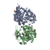
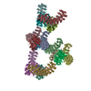
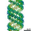
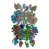
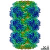
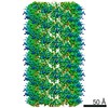
| Title | EM structure of Bacillus subtilis ribonucleotide reductase inhibited double-helical filament of NrdE alpha subunit with dATP | |||||||||
 Components Components | Ribonucleoside-diphosphate reductase | |||||||||
 Keywords Keywords | OXIDOREDUCTASE / PROTEIN FIBRIL / ribonucleotide reductase / allostery / nucleotide metabolism / filament / dATP / ATP | |||||||||
| Function / homology |  Function and homology information Function and homology informationribonucleoside-diphosphate reductase complex / ribonucleoside-diphosphate reductase / ribonucleoside-diphosphate reductase activity, thioredoxin disulfide as acceptor / deoxyribonucleotide biosynthetic process / DNA replication / ATP binding Similarity search - Function | |||||||||
| Biological species |  | |||||||||
| Method | ELECTRON MICROSCOPY / helical reconstruction / cryo EM / Resolution: 6 Å | |||||||||
 Authors Authors | Thomas, W.C. / Bacik, J.P. / Chen, J.Z. / Ando, N. | |||||||||
| Funding support |  United States, 2items United States, 2items
| |||||||||
 Citation Citation | Journal: Nat Commun / Year: 2019 Title: Convergent allostery in ribonucleotide reductase. Authors: William C Thomas / F Phil Brooks / Audrey A Burnim / John-Paul Bacik / JoAnne Stubbe / Jason T Kaelber / James Z Chen / Nozomi Ando / Abstract: Ribonucleotide reductases (RNRs) use a conserved radical-based mechanism to catalyze the conversion of ribonucleotides to deoxyribonucleotides. Within the RNR family, class Ib RNRs are notable for ...Ribonucleotide reductases (RNRs) use a conserved radical-based mechanism to catalyze the conversion of ribonucleotides to deoxyribonucleotides. Within the RNR family, class Ib RNRs are notable for being largely restricted to bacteria, including many pathogens, and for lacking an evolutionarily mobile ATP-cone domain that allosterically controls overall activity. In this study, we report the emergence of a distinct and unexpected mechanism of activity regulation in the sole RNR of the model organism Bacillus subtilis. Using a hypothesis-driven structural approach that combines the strengths of small-angle X-ray scattering (SAXS), crystallography, and cryo-electron microscopy (cryo-EM), we describe the reversible interconversion of six unique structures, including a flexible active tetramer and two inhibited helical filaments. These structures reveal the conformational gymnastics necessary for RNR activity and the molecular basis for its control via an evolutionarily convergent form of allostery. | |||||||||
| History |
|
- Structure visualization
| Movie |
Movie viewer |
|---|---|
| Structure viewer | Molecule: MolmilJmol/JSmol |
- Downloads & links
-Download
| PDBx/mmCIF format | 6myx.cif.gz | 477.4 KB | Display | PDBx/mmCIF format |
|---|---|---|---|---|
| PDB format | pdb6myx.ent.gz | 397 KB | Display | PDB format |
| PDBx/mmJSON format | 6myx.json.gz | Tree view | PDBx/mmJSON format | |
| Others |  Other downloads Other downloads |
-Validation report
| Arichive directory | https://data.pdbj.org/pub/pdb/validation_reports/my/6myxftp://data.pdbj.org/pub/pdb/validation_reports/my/6myx | HTTPS FTP |
|---|
-Related structure data
| Related structure data | 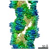 9293MC 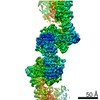 9272C 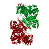 6mt9C 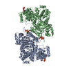 6mv9C 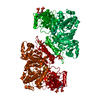 6mveC 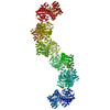 6mw3C C: citing same article ( M: map data used to model this data |
|---|---|
| Similar structure data |
-Links
 PDBj
PDBj


- Assembly
| Deposited unit | 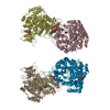
|
|---|---|
| 1 | 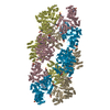
|
| 2 |
|
| Symmetry | Helical symmetry: (Circular symmetry: 1 / Dyad axis: no / N subunits divisor: 1 / Num. of operations: 3 / Rise per n subunits: 74.24 Å / Rotation per n subunits: 81.28 °) |
-Components
| #1: Protein | Mass: 80791.469 Da / Num. of mol.: 4 Source method: isolated from a genetically manipulated source Source: (gene. exp.) References: UniProt: A0A162Q3J9, UniProt: P50620*PLUS, ribonucleoside-diphosphate reductase #2: Chemical | ChemComp-DTP /   Mass: 491.182 Da / Num. of mol.: 8 / Source method: obtained synthetically / Formula: C10H16N5O12P3 Mass: 491.182 Da / Num. of mol.: 8 / Source method: obtained synthetically / Formula: C10H16N5O12P3 |
|---|
-Experimental details
-Experiment
| Experiment | Method: ELECTRON MICROSCOPY |
|---|---|
| EM experiment | Aggregation state: FILAMENT / 3D reconstruction method: helical reconstruction |
- Sample preparation
| Component | Name: Inhibited filament of ribonucleoside-diphosphate reductase composed of NrdE alpha subunit Type: COMPLEX Details: The filament is a double helix. Each helix is composed of NrdE subunits dimerizing at alternating canonical and non-canonical interfaces. Entity ID: #1 / Source: RECOMBINANT | |||||||||||||||||||||||||
|---|---|---|---|---|---|---|---|---|---|---|---|---|---|---|---|---|---|---|---|---|---|---|---|---|---|---|
| Molecular weight | Experimental value: NO | |||||||||||||||||||||||||
| Source (natural) | Organism: | |||||||||||||||||||||||||
| Source (recombinant) | Organism: | |||||||||||||||||||||||||
| Buffer solution | pH: 7.6 Details: Glycerol in original buffer was diluted to < 0.25% w/v. | |||||||||||||||||||||||||
| Buffer component |
| |||||||||||||||||||||||||
| Specimen | Conc.: 0.81 mg/ml / Embedding applied: NO / Shadowing applied: NO / Staining applied: NO / Vitrification applied: YES Details: Samples of the dATP-induced NrdE filament were produced by incubating 40 uM holo-NrdE with 100 uM dATP and 1 mM CDP in assay buffer. The mixture was then diluted to 10 uM NrdE in the same ...Details: Samples of the dATP-induced NrdE filament were produced by incubating 40 uM holo-NrdE with 100 uM dATP and 1 mM CDP in assay buffer. The mixture was then diluted to 10 uM NrdE in the same nucleotide-containing buffer. | |||||||||||||||||||||||||
| Specimen support | Grid material: COPPER / Grid mesh size: 300 divisions/in. / Grid type: Quantifoil R1.2/1.3 | |||||||||||||||||||||||||
| Vitrification | Instrument: FEI VITROBOT MARK IV / Cryogen name: ETHANE / Humidity: 90 % / Chamber temperature: 300 K / Details: 3.5 seconds blotting |
- Electron microscopy imaging
| Experimental equipment |  Model: Talos Arctica / Image courtesy: FEI Company |
|---|---|
| Microscopy | Model: FEI TALOS ARCTICA |
| Electron gun | Electron source:  FIELD EMISSION GUN / Accelerating voltage: 200 kV / Illumination mode: FLOOD BEAM FIELD EMISSION GUN / Accelerating voltage: 200 kV / Illumination mode: FLOOD BEAM |
| Electron lens | Mode: BRIGHT FIELD / Nominal defocus max: 3000 nm / Nominal defocus min: 1200 nm / Calibrated defocus min: 1200 nm / Calibrated defocus max: 3000 nm / Cs: 2.7 mm / C2 aperture diameter: 70 µm |
| Specimen holder | Cryogen: NITROGEN / Specimen holder model: FEI TITAN KRIOS AUTOGRID HOLDER |
| Image recording | Average exposure time: 20 sec. / Electron dose: 20 e/Å2 / Detector mode: COUNTING / Film or detector model: GATAN K2 SUMMIT (4k x 4k) / Num. of grids imaged: 2 / Num. of real images: 500 |
| EM imaging optics | Chromatic aberration corrector: None / Spherical aberration corrector: None |
| Image scans | Sampling size: 5 µm / Width: 4000 / Height: 4000 / Movie frames/image: 100 / Used frames/image: 2-90 |
- Processing
| Software | Name: PHENIX / Version: 1.14_3260: / Classification: refinement | |||||||||||||||||||||
|---|---|---|---|---|---|---|---|---|---|---|---|---|---|---|---|---|---|---|---|---|---|---|
| EM software |
| |||||||||||||||||||||
| CTF correction | Type: PHASE FLIPPING AND AMPLITUDE CORRECTION | |||||||||||||||||||||
| Helical symmerty | Angular rotation/subunit: 81.28 ° / Axial rise/subunit: 74.24 Å / Axial symmetry: C1 | |||||||||||||||||||||
| 3D reconstruction | Resolution: 6 Å / Resolution method: FSC 0.143 CUT-OFF / Num. of particles: 4 / Symmetry type: HELICAL | |||||||||||||||||||||
| Atomic model building | Protocol: OTHER / Space: REAL / Target criteria: Corellation coefficient | |||||||||||||||||||||
| Atomic model building |
|