 Movie
Movie Controller

[English] 日本語
 Yorodumi
- EMDB-9272: EM structure of Bacillus subtilis ribonucleotide reductase inhibi... -
+ Open data

- Basic information
| Entry | Database: EMDB / ID: EMD-9272 | |||||||||
|---|---|---|---|---|---|---|---|---|---|---|
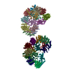
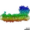
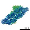
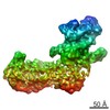
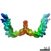
| Title | EM structure of Bacillus subtilis ribonucleotide reductase inhibited filament composed of NrdE alpha subunit and NrdF beta subunit with dATP | |||||||||
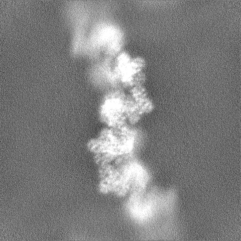
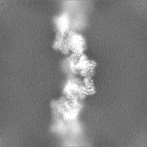
 Map data Map data | ASU of Bacillus subtilis ribonucleotide reductase inhibited filament composed of NrdE alpha and NrdF beta subunits with dATP. Handedness already corrected by reference to NrdE crystal structures. | |||||||||
 Sample Sample |
| |||||||||
 Keywords Keywords | ribonucleotide reductase / allostery / nucleotide metabolism / filament / dATP / ATP / OXIDOREDUCTASE | |||||||||
| Function / homology |  Function and homology information Function and homology informationribonucleoside-diphosphate reductase complex / ribonucleoside-diphosphate reductase / ribonucleoside-diphosphate reductase activity, thioredoxin disulfide as acceptor / deoxyribonucleotide biosynthetic process / DNA replication / ATP binding Similarity search - Function | |||||||||
| Biological species |  | |||||||||
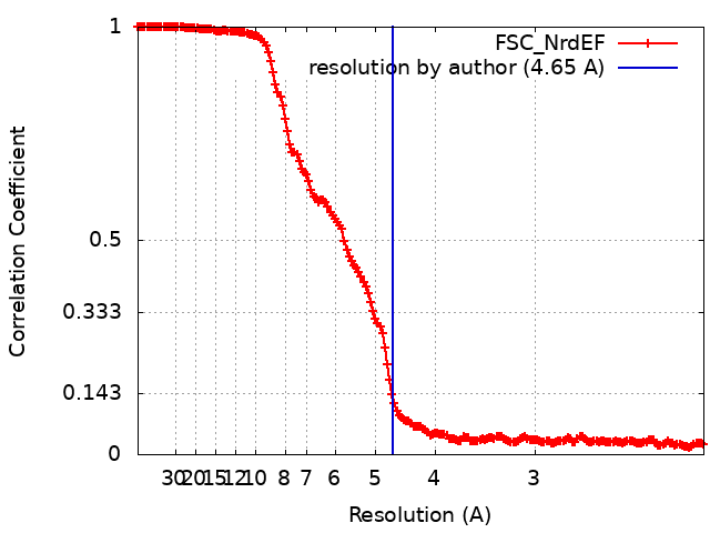
| Method | helical reconstruction / cryo EM / Resolution: 4.65 Å | |||||||||
 Authors Authors | Thomas WC / Bacik JP / Kaelber JT / Ando N | |||||||||
| Funding support |  United States, 2 items United States, 2 items
| |||||||||
 Citation Citation | Journal: Nat Commun / Year: 2019 Title: Convergent allostery in ribonucleotide reductase. Authors: William C Thomas / F Phil Brooks / Audrey A Burnim / John-Paul Bacik / JoAnne Stubbe / Jason T Kaelber / James Z Chen / Nozomi Ando / Abstract: Ribonucleotide reductases (RNRs) use a conserved radical-based mechanism to catalyze the conversion of ribonucleotides to deoxyribonucleotides. Within the RNR family, class Ib RNRs are notable for ...Ribonucleotide reductases (RNRs) use a conserved radical-based mechanism to catalyze the conversion of ribonucleotides to deoxyribonucleotides. Within the RNR family, class Ib RNRs are notable for being largely restricted to bacteria, including many pathogens, and for lacking an evolutionarily mobile ATP-cone domain that allosterically controls overall activity. In this study, we report the emergence of a distinct and unexpected mechanism of activity regulation in the sole RNR of the model organism Bacillus subtilis. Using a hypothesis-driven structural approach that combines the strengths of small-angle X-ray scattering (SAXS), crystallography, and cryo-electron microscopy (cryo-EM), we describe the reversible interconversion of six unique structures, including a flexible active tetramer and two inhibited helical filaments. These structures reveal the conformational gymnastics necessary for RNR activity and the molecular basis for its control via an evolutionarily convergent form of allostery. | |||||||||
| History |
|
- Structure visualization
| Movie |
Movie viewer |
|---|---|
| Structure viewer | EM map: SurfViewMolmilJmol/JSmol |
| Supplemental images |

- Downloads & links
-EMDB archive
| Map data | emd_9272.map.gz | 9.7 MB | EMDB map data format | |
|---|---|---|---|---|
| Header (meta data) | emd-9272-v30.xmlemd-9272.xml | 21.2 KB 21.2 KB | Display Display | EMDB header |
| FSC (resolution estimation) | emd_9272_fsc.xml | 16.8 KB | Display | FSC data file |
| Images | 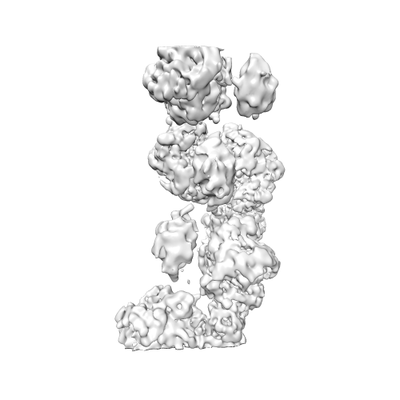 emd_9272.png emd_9272.png | 31.9 KB | ||
| Masks | emd_9272_msk_1.map | 421.9 MB | Mask map | |
| Filedesc metadata | emd-9272.cif.gz | 7.5 KB | ||
| Others | emd_9272_half_map_1.map.gzemd_9272_half_map_2.map.gz | 391.2 MB 391.2 MB | ||
| Archive directory |  http://ftp.pdbj.org/pub/emdb/structures/EMD-9272ftp://ftp.pdbj.org/pub/emdb/structures/EMD-9272 http://ftp.pdbj.org/pub/emdb/structures/EMD-9272ftp://ftp.pdbj.org/pub/emdb/structures/EMD-9272 | HTTPS FTP |
-Related structure data
| Related structure data | 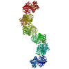 6mw3MC 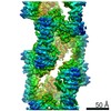 9293C 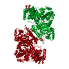 6mt9C 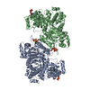 6mv9C 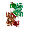 6mveC  6myxC C: citing same article ( M: atomic model generated by this map |
|---|---|
| Similar structure data |

-Links
| EMDB pages | EMDB (EBI/PDBe) / EMDataResource |
|---|---|
| Related items in Molecule of the Month |

-Map
| File | Download / File: emd_9272.map.gz / Format: CCP4 / Size: 52.7 MB / Type: IMAGE STORED AS FLOATING POINT NUMBER (4 BYTES) | ||||||||||||||||||||||||||||||||||||||||||||||||||||||||||||||||||||
|---|---|---|---|---|---|---|---|---|---|---|---|---|---|---|---|---|---|---|---|---|---|---|---|---|---|---|---|---|---|---|---|---|---|---|---|---|---|---|---|---|---|---|---|---|---|---|---|---|---|---|---|---|---|---|---|---|---|---|---|---|---|---|---|---|---|---|---|---|---|
| Annotation | ASU of Bacillus subtilis ribonucleotide reductase inhibited filament composed of NrdE alpha and NrdF beta subunits with dATP. Handedness already corrected by reference to NrdE crystal structures. | ||||||||||||||||||||||||||||||||||||||||||||||||||||||||||||||||||||
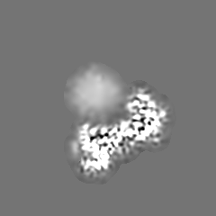
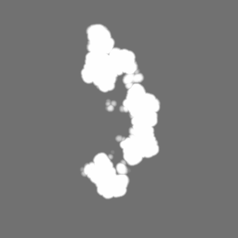
| Projections & slices | Image control
Images are generated by Spider. | ||||||||||||||||||||||||||||||||||||||||||||||||||||||||||||||||||||
| Voxel size | X=Y=Z: 1.05 Å | ||||||||||||||||||||||||||||||||||||||||||||||||||||||||||||||||||||
| Density |
| ||||||||||||||||||||||||||||||||||||||||||||||||||||||||||||||||||||
| Symmetry | Space group: 1 | ||||||||||||||||||||||||||||||||||||||||||||||||||||||||||||||||||||
| Details | EMDB XML:
CCP4 map header:
| ||||||||||||||||||||||||||||||||||||||||||||||||||||||||||||||||||||
 Z (Sec.)
 Y (Row.)
 X (Col.)


-Supplemental data
-Mask #1
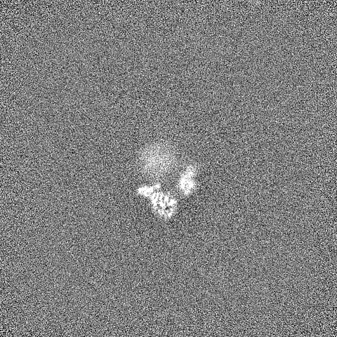
| File | emd_9272_msk_1.map | ||||||||||||
|---|---|---|---|---|---|---|---|---|---|---|---|---|---|
| Projections & Slices |
| ||||||||||||
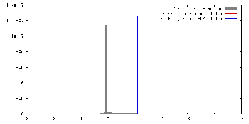
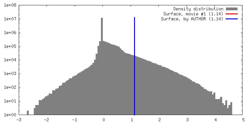
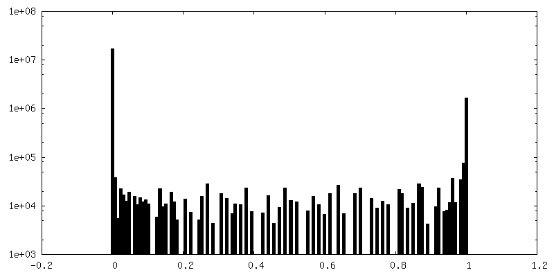
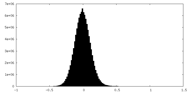
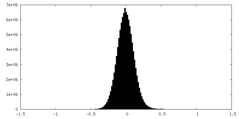
| Density Histograms |


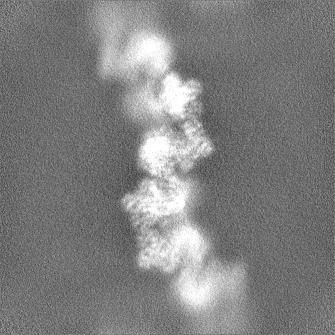
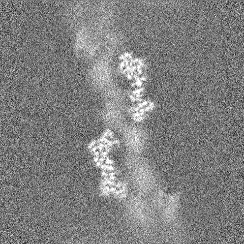
-Half map: Unfiltered, unmasked, half-map from refinement of NrdEF
| File | emd_9272_half_map_1.map | ||||||||||||
|---|---|---|---|---|---|---|---|---|---|---|---|---|---|
| Annotation | Unfiltered, unmasked, half-map from refinement of NrdEF | ||||||||||||
| Projections & Slices |
| ||||||||||||
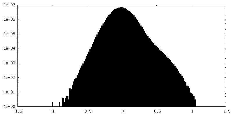
| Density Histograms |


-Half map: Unfiltered, unmasked half-map from refinement of NrdEF
| File | emd_9272_half_map_2.map | ||||||||||||
|---|---|---|---|---|---|---|---|---|---|---|---|---|---|
| Annotation | Unfiltered, unmasked half-map from refinement of NrdEF | ||||||||||||
| Projections & Slices |
| ||||||||||||
| Density Histograms |


- Sample components
-Entire : Inhibited filament of ribonucleoside-diphosphate reductase compos...
| Entire | Name: Inhibited filament of ribonucleoside-diphosphate reductase composed of NrdE alpha subunits and NrdF beta subunit tails |
|---|---|
| Components |
|
-Supramolecule #1: Inhibited filament of ribonucleoside-diphosphate reductase compos...
| Supramolecule | Name: Inhibited filament of ribonucleoside-diphosphate reductase composed of NrdE alpha subunits and NrdF beta subunit tails type: complex / ID: 1 / Parent: 0 / Macromolecule list: #1-#2 Details: Beta subunit core density only visible at low threshold. Beta subunit tail is bound with strong density to alpha subunit and modeled as a poly-A peptide in the model. |
|---|---|
| Source (natural) | Organism: |
-Macromolecule #1: Ribonucleoside-diphosphate reductase
| Macromolecule | Name: Ribonucleoside-diphosphate reductase / type: protein_or_peptide / ID: 1 / Number of copies: 2 / Enantiomer: LEVO / EC number: ribonucleoside-diphosphate reductase |
|---|---|
| Source (natural) | Organism: |
| Molecular weight | Theoretical: 80.791469 KDa |
| Recombinant expression | Organism: |
| Sequence | String: MSQNQVPKWI QLNNEIMIQK DGKFQFDKDK EAVHSYFVDY INQNTVFFHN LKEKLDYLVE NQYYEEEFLS LYSFEDIKEV FKTAYAKKF RFPSFMSAFK FYNDYALKTN DKKKILERYE DRISIVALFF ANGDTEKAKE YVNLMINQEY QPSTPTFLNA G RKRRGELV ...String: MSQNQVPKWI QLNNEIMIQK DGKFQFDKDK EAVHSYFVDY INQNTVFFHN LKEKLDYLVE NQYYEEEFLS LYSFEDIKEV FKTAYAKKF RFPSFMSAFK FYNDYALKTN DKKKILERYE DRISIVALFF ANGDTEKAKE YVNLMINQEY QPSTPTFLNA G RKRRGELV SCFLLEVNDS LNDISRAIDI SMQLSKLGGG VSLNLSKLRA KGEAIKDVEN ATKGVVGVMK LLDNAFRYAD QM GQRQGSG AAYLNIFHRD INDFLDTKKI SADEDVRVKT LSIGVVIPDK FVELAREDKA AYVFYPHTIY KEYGQHMDEM DMN EMYDKF VDNPRVKKEK INPRKLLEKL AMLRSESGYP YIMFQDNVNK VHANNHISKV KFSNLCSEVL QASQVSSYTD YDEE DEIGL DISCNLGSLN ILNVMEHKSI EKTVKLATDS LTHVSETTDI RNAPAVRRAN KAMKSIGLGA MNLHGYLAQN GIAYE SPEA RDFANTFFMM VNFYSIQRSA EIAKEKGETF DQYEGSTYAT GEYFDKYVST DFSPKYEKIA NLFEGMHIPT TEDWKK LKA FVAEHGMYHS YRLCIAPTGS ISYVQSSTAS VMPIMERIEE RTYGNSKTYY PMPGLASNNW FFYKEAYDMD MFKVVDM IA TIQQHIDQGI SFTLFLKDTM TTRDLNRIDL YAHHRGIKTI YYARTKDTGQ DSCLSCVV UniProtKB: Ribonucleoside-diphosphate reductase |
-Macromolecule #2: Ribonucleoside-diphosphate reductase NrdF beta subunit
| Macromolecule | Name: Ribonucleoside-diphosphate reductase NrdF beta subunit type: protein_or_peptide / ID: 2 / Details: C-terminus modeled as a polyalanine chain / Number of copies: 2 / Enantiomer: LEVO |
|---|---|
| Source (natural) | Organism: |
| Molecular weight | Theoretical: 698.854 Da |
| Recombinant expression | Organism: |
| Sequence | String: (UNK)(UNK)(UNK)(UNK)(UNK)(UNK)(UNK)(UNK) |
-Macromolecule #3: 2'-DEOXYADENOSINE 5'-TRIPHOSPHATE
| Macromolecule | Name: 2'-DEOXYADENOSINE 5'-TRIPHOSPHATE / type: ligand / ID: 3 / Number of copies: 4 / Formula: DTP |
|---|---|
| Molecular weight | Theoretical: 491.182 Da |
| Chemical component information |  ChemComp-DTP: |
-Experimental details
-Structure determination
| Method | cryo EM |
|---|---|
 Processing Processing | helical reconstruction |
| Aggregation state | filament |
-Sample preparation
| Concentration | 0.40 mg/mL | |||||||||||||||
|---|---|---|---|---|---|---|---|---|---|---|---|---|---|---|---|---|
| Buffer | pH: 7.6 Component:
Details: Glycerol in original storage buffer was diluted to < 0.25% w/v. | |||||||||||||||
| Grid | Details: unspecified | |||||||||||||||
| Vitrification | Cryogen name: ETHANE / Chamber humidity: 95 % / Instrument: LEICA EM GP | |||||||||||||||
| Details | Cryo-EM samples of the NrdEF filament were prepared by mixing 20 uM C382S holo-NrdE with 20 or 40 uM Mn-reconstituted NrdF in assay buffer with 100 uM dATP and 1 mM CDP, prior to dilution with nucleotide-containing buffer to a concentration of 5 uM protein. A subset of the grids were pre-coated with a support film of continuous, amorphous carbon by flotation of cleaved mica. For these grids, the sample was diluted to a final protein concentration of 2 uM. |
- Electron microscopy
| Microscope | FEI TALOS ARCTICA |
|---|---|
| Specialist optics | Energy filter - Slit width: 20 eV |
| Image recording | Film or detector model: GATAN K2 SUMMIT (4k x 4k) / Detector mode: COUNTING / Digitization - Dimensions - Width: 3838 pixel / Digitization - Dimensions - Height: 3710 pixel / Number real images: 2843 / Average electron dose: 8.0 e/Å2 |
| Electron beam | Acceleration voltage: 200 kV / Electron source:  FIELD EMISSION GUN FIELD EMISSION GUN |
| Electron optics | C2 aperture diameter: 50.0 µm / Illumination mode: FLOOD BEAM / Imaging mode: BRIGHT FIELD / Cs: 2.7 mm / Nominal magnification: 130000 |
| Sample stage | Specimen holder model: FEI TITAN KRIOS AUTOGRID HOLDER / Cooling holder cryogen: NITROGEN |
| Experimental equipment |  Model: Talos Arctica / Image courtesy: FEI Company |
+Image processing
-Atomic model buiding 1
| Initial model | PDB ID: Chain - Source name: PDB / Chain - Initial model type: experimental model |
|---|---|
| Refinement | Space: REAL / Protocol: OTHER / Target criteria: Correlation coefficient |
| Output model | PDB-6mw3: |

