 Movie
Movie Controller

[English] 日本語
 Yorodumi
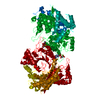
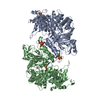
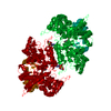
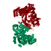
- PDB-6mt9: X-ray crystal structure of Bacillus subtilis ribonucleotide reduc... -
+ Open data

- Basic information
| Entry | Database: PDB / ID: 6mt9 | |||||||||
|---|---|---|---|---|---|---|---|---|---|---|
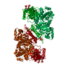
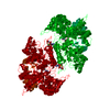
| Title | X-ray crystal structure of Bacillus subtilis ribonucleotide reductase NrdE alpha subunit with TTP, ATP, and ADP | |||||||||
 Components Components | Ribonucleoside-diphosphate reductase | |||||||||
 Keywords Keywords | OXIDOREDUCTASE / ribonucleotide reductase / allostery / nucleotide metabolism / dATP / ATP | |||||||||
| Function / homology |  Function and homology information Function and homology informationribonucleoside-diphosphate reductase complex / ribonucleoside-diphosphate reductase / ribonucleoside-diphosphate reductase activity, thioredoxin disulfide as acceptor / deoxyribonucleotide biosynthetic process / DNA replication / ATP binding Similarity search - Function | |||||||||
| Biological species |  | |||||||||
| Method |  X-RAY DIFFRACTION / SYNCHROTRON / MOLECULAR REPLACEMENT / Resolution: 2.5 Å X-RAY DIFFRACTION / SYNCHROTRON / MOLECULAR REPLACEMENT / Resolution: 2.5 Å | |||||||||
 Authors Authors | Thomas, W.C. / Brooks, F.P. / Bacik, J.P. / Ando, N. | |||||||||
| Funding support |  United States, 2items United States, 2items
| |||||||||
 Citation Citation | Journal: Nat Commun / Year: 2019 Title: Convergent allostery in ribonucleotide reductase. Authors: William C Thomas / F Phil Brooks / Audrey A Burnim / John-Paul Bacik / JoAnne Stubbe / Jason T Kaelber / James Z Chen / Nozomi Ando / Abstract: Ribonucleotide reductases (RNRs) use a conserved radical-based mechanism to catalyze the conversion of ribonucleotides to deoxyribonucleotides. Within the RNR family, class Ib RNRs are notable for ...Ribonucleotide reductases (RNRs) use a conserved radical-based mechanism to catalyze the conversion of ribonucleotides to deoxyribonucleotides. Within the RNR family, class Ib RNRs are notable for being largely restricted to bacteria, including many pathogens, and for lacking an evolutionarily mobile ATP-cone domain that allosterically controls overall activity. In this study, we report the emergence of a distinct and unexpected mechanism of activity regulation in the sole RNR of the model organism Bacillus subtilis. Using a hypothesis-driven structural approach that combines the strengths of small-angle X-ray scattering (SAXS), crystallography, and cryo-electron microscopy (cryo-EM), we describe the reversible interconversion of six unique structures, including a flexible active tetramer and two inhibited helical filaments. These structures reveal the conformational gymnastics necessary for RNR activity and the molecular basis for its control via an evolutionarily convergent form of allostery. | |||||||||
| History |
|
- Structure visualization
| Structure viewer | Molecule: MolmilJmol/JSmol |
|---|
- Downloads & links
-Download
| PDBx/mmCIF format | 6mt9.cif.gz | 159.1 KB | Display | PDBx/mmCIF format |
|---|---|---|---|---|
| PDB format | pdb6mt9.ent.gz | 120.9 KB | Display | PDB format |
| PDBx/mmJSON format | 6mt9.json.gz | Tree view | PDBx/mmJSON format | |
| Others |  Other downloads Other downloads |
-Validation report
| Arichive directory | https://data.pdbj.org/pub/pdb/validation_reports/mt/6mt9ftp://data.pdbj.org/pub/pdb/validation_reports/mt/6mt9 | HTTPS FTP |
|---|
-Related structure data
| Related structure data | 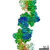 9272C 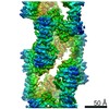 9293C 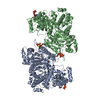 6mv9C  6mveC 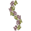 6mw3C 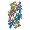 6myxC 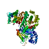 6cgmS C: citing same article ( S: Starting model for refinement |
|---|---|
| Similar structure data |


-Links
 PDBj
PDBj
- Assembly
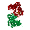
| Deposited unit | 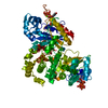
| ||||||||
|---|---|---|---|---|---|---|---|---|---|
| 1 | 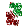
| ||||||||
| Unit cell |
|
-Components
-Protein , 1 types, 1 molecules A
| #1: Protein | Mass: 80791.469 Da / Num. of mol.: 1 Source method: isolated from a genetically manipulated source Source: (gene. exp.) References: UniProt: A0A162Q3J9, UniProt: P50620*PLUS, ribonucleoside-diphosphate reductase |
|---|
-Non-polymers , 5 types, 198 molecules 


| #2: Chemical | ChemComp-TTP /  Mass: 482.168 Da / Num. of mol.: 1 / Source method: obtained synthetically / Formula: C10H17N2O14P3 Mass: 482.168 Da / Num. of mol.: 1 / Source method: obtained synthetically / Formula: C10H17N2O14P3 |
|---|---|
| #3: Chemical | ChemComp-ADP /  Mass: 427.201 Da / Num. of mol.: 1 / Source method: obtained synthetically / Formula: C10H15N5O10P2 / Comment: ADP, energy-carrying molecule*YM Mass: 427.201 Da / Num. of mol.: 1 / Source method: obtained synthetically / Formula: C10H15N5O10P2 / Comment: ADP, energy-carrying molecule*YM |
| #4: Chemical | ChemComp-ATP /  Mass: 507.181 Da / Num. of mol.: 1 / Source method: obtained synthetically / Formula: C10H16N5O13P3 / Comment: ATP, energy-carrying molecule*YM Mass: 507.181 Da / Num. of mol.: 1 / Source method: obtained synthetically / Formula: C10H16N5O13P3 / Comment: ATP, energy-carrying molecule*YM |
| #5: Chemical | ChemComp-MG /  Mass: 24.305 Da / Num. of mol.: 1 / Source method: obtained synthetically / Formula: Mg Mass: 24.305 Da / Num. of mol.: 1 / Source method: obtained synthetically / Formula: Mg |
| #6: Water | ChemComp-HOH / Mass: 18.015 Da / Num. of mol.: 194 / Source method: isolated from a natural source / Formula: H2O |
-Details
| Has protein modification | Y |
|---|
-Experimental details
-Experiment
| Experiment | Method: X-RAY DIFFRACTION / Number of used crystals: 1 |
|---|
- Sample preparation
| Crystal | Density Matthews: 3.1 Å3/Da / Density % sol: 60.29 % |
|---|---|
| Crystal grow | Temperature: 293 K / Method: vapor diffusion, sitting drop / pH: 7.6 Details: The crystal was grown by vapor diffusion from 4.5 mg/ml holo-NrdE in 50 mM HEPES (pH 7.6), 50 mM NaCl, 5 mM MgCl2, 2 mM TCEP, and 1% glycerol, supplemented with 5 mM ATP, 0.5 mM TTP, and 1 ...Details: The crystal was grown by vapor diffusion from 4.5 mg/ml holo-NrdE in 50 mM HEPES (pH 7.6), 50 mM NaCl, 5 mM MgCl2, 2 mM TCEP, and 1% glycerol, supplemented with 5 mM ATP, 0.5 mM TTP, and 1 mM GDP. The protein solution was incubated for 10 minutes with freshly added nucleotides prior to being mixed in a 1:1 hanging drop with a precipitating solution of 6% PEG 3350, 1% w/v tryptone, and 50 mM HEPES at pH 7. Crystals were cryoprotected by soaking for 5-10 seconds in well solution mixed with 8% w/v sucrose, 2% w/v glucose, 8% v/v glycerol, 8% v/v ethylene glycol and supplemented with nucleotides, TCEP, and MgCl2 adjusted to the same concentrations used in the original protein solutions. |
-Data collection
| Diffraction | Mean temperature: 100 K / Serial crystal experiment: N |
|---|---|
| Diffraction source | Source: SYNCHROTRON / Site: CHESS / Beamline: F1 / Wavelength: 0.9775 Å |
| Detector | Type: DECTRIS PILATUS 6M / Detector: PIXEL / Date: May 5, 2018 |
| Radiation | Protocol: SINGLE WAVELENGTH / Monochromatic (M) / Laue (L): M / Scattering type: x-ray |
| Radiation wavelength | Wavelength: 0.9775 Å / Relative weight: 1 |
| Reflection | Resolution: 2.5→16 Å / Num. obs: 35371 / % possible obs: 99.2 % / Redundancy: 17 % / CC1/2: 0.998 / Rmerge(I) obs: 0.175 / Rpim(I) all: 0.041 / Net I/σ(I): 14.7 |
| Reflection shell | Resolution: 2.5→2.6 Å / Redundancy: 17.6 % / Rmerge(I) obs: 1.445 / Mean I/σ(I) obs: 2.2 / Num. unique obs: 3958 / CC1/2: 0.734 / Rpim(I) all: 0.333 / % possible all: 99.9 |
- Processing
| Software |
| |||||||||||||||||||||||||||||||||||||||||||||||||||||||||||||||||||||||||||||||||||||||||||||||||||||||||||||||||||||||||||||||||||||||||||||||||||
|---|---|---|---|---|---|---|---|---|---|---|---|---|---|---|---|---|---|---|---|---|---|---|---|---|---|---|---|---|---|---|---|---|---|---|---|---|---|---|---|---|---|---|---|---|---|---|---|---|---|---|---|---|---|---|---|---|---|---|---|---|---|---|---|---|---|---|---|---|---|---|---|---|---|---|---|---|---|---|---|---|---|---|---|---|---|---|---|---|---|---|---|---|---|---|---|---|---|---|---|---|---|---|---|---|---|---|---|---|---|---|---|---|---|---|---|---|---|---|---|---|---|---|---|---|---|---|---|---|---|---|---|---|---|---|---|---|---|---|---|---|---|---|---|---|---|---|---|---|
| Refinement | Method to determine structure: MOLECULAR REPLACEMENT Starting model: 6CGM Resolution: 2.5→15.998 Å / SU ML: 0.26 / Cross valid method: FREE R-VALUE / σ(F): 1.33 / Phase error: 21.99
| |||||||||||||||||||||||||||||||||||||||||||||||||||||||||||||||||||||||||||||||||||||||||||||||||||||||||||||||||||||||||||||||||||||||||||||||||||
| Solvent computation | Shrinkage radii: 0.9 Å / VDW probe radii: 1.11 Å | |||||||||||||||||||||||||||||||||||||||||||||||||||||||||||||||||||||||||||||||||||||||||||||||||||||||||||||||||||||||||||||||||||||||||||||||||||
| Refinement step | Cycle: LAST / Resolution: 2.5→15.998 Å
| |||||||||||||||||||||||||||||||||||||||||||||||||||||||||||||||||||||||||||||||||||||||||||||||||||||||||||||||||||||||||||||||||||||||||||||||||||
| Refine LS restraints |
| |||||||||||||||||||||||||||||||||||||||||||||||||||||||||||||||||||||||||||||||||||||||||||||||||||||||||||||||||||||||||||||||||||||||||||||||||||
| LS refinement shell |
|
