 Movie
Movie Controller


+ Open data

- Basic information
| Entry | Database: PDB / ID: 2cvt | ||||||
|---|---|---|---|---|---|---|---|
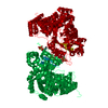
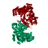
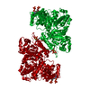
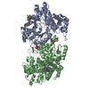
| Title | Structures of Yeast Ribonucleotide Reductase I | ||||||
 Components Components | Ribonucleoside-diphosphate reductase large chain 1 | ||||||
 Keywords Keywords | OXIDOREDUCTASE / Eukaryotic / Ribonucleotide Reductase / dNTP Regulation | ||||||
| Function / homology |  Function and homology information Function and homology informationribonucleoside-diphosphate reductase complex / ribonucleoside-diphosphate reductase / ribonucleoside-diphosphate reductase activity, thioredoxin disulfide as acceptor / deoxyribonucleotide biosynthetic process / nucleotide binding / ATP binding / identical protein binding / nucleus / cytoplasm Similarity search - Function | ||||||
| Biological species |  | ||||||
| Method |  X-RAY DIFFRACTION / SYNCHROTRON / MOLECULAR REPLACEMENT / Resolution: 3.2 Å X-RAY DIFFRACTION / SYNCHROTRON / MOLECULAR REPLACEMENT / Resolution: 3.2 Å | ||||||
 Authors Authors | Xu, H. / Faber, C. / Uchiki, T. / Fairman, J.W. / Racca, J. / Dealwis, C. | ||||||
 Citation Citation | Journal: Proc.Natl.Acad.Sci.Usa / Year: 2006 Title: Structures of eukaryotic ribonucleotide reductase I provide insights into dNTP regulation Authors: Xu, H. / Faber, C. / Uchiki, T. / Fairman, J.W. / Racca, J. / Dealwis, C. | ||||||
| History |
|
- Structure visualization
| Structure viewer | Molecule: MolmilJmol/JSmol |
|---|
- Downloads & links
-Download
| PDBx/mmCIF format | 2cvt.cif.gz | 145 KB | Display | PDBx/mmCIF format |
|---|---|---|---|---|
| PDB format | pdb2cvt.ent.gz | 109.9 KB | Display | PDB format |
| PDBx/mmJSON format | 2cvt.json.gz | Tree view | PDBx/mmJSON format | |
| Others |  Other downloads Other downloads |
-Validation report
| Arichive directory | https://data.pdbj.org/pub/pdb/validation_reports/cv/2cvtftp://data.pdbj.org/pub/pdb/validation_reports/cv/2cvt | HTTPS FTP |
|---|
-Related structure data
| Related structure data |  1zyzC  1zzdC  2cvsC  2cvuC  2cvvC  2cvwC  2cvxC  2cvyC C: citing same article ( |
|---|---|
| Similar structure data |


-Links
 PDBj
PDBj


- Assembly
| Deposited unit | 
| ||||||||
|---|---|---|---|---|---|---|---|---|---|
| 1 | 
| ||||||||
| Unit cell |
| ||||||||
| Details | The second part of the active biological dimer assembly is generated by the two fold axis: -x, -y+1, z. |
-Components
| #1: Protein | Mass: 99672.984 Da / Num. of mol.: 1 Source method: isolated from a genetically manipulated source Source: (gene. exp.) Plasmid: pWJ751-3 / Species (production host): Escherichia coli / Production host:  References: UniProt: P21524, ribonucleoside-diphosphate reductase |
|---|---|
| #2: Chemical | ChemComp-MG /   Mass: 24.305 Da / Num. of mol.: 1 / Source method: obtained synthetically / Formula: Mg Mass: 24.305 Da / Num. of mol.: 1 / Source method: obtained synthetically / Formula: Mg |
| #3: Chemical | ChemComp-ANP /   Mass: 506.196 Da / Num. of mol.: 1 / Source method: obtained synthetically / Formula: C10H17N6O12P3 / Comment: AMP-PNP, energy-carrying molecule analogue*YM Mass: 506.196 Da / Num. of mol.: 1 / Source method: obtained synthetically / Formula: C10H17N6O12P3 / Comment: AMP-PNP, energy-carrying molecule analogue*YM |
-Experimental details
-Experiment
| Experiment | Method: X-RAY DIFFRACTION / Number of used crystals: 1 |
|---|
- Sample preparation
| Crystal | Density Matthews: 2.04 Å3/Da / Density % sol: 37.3 % |
|---|---|
| Crystal grow | Temperature: 298 K / Method: evaporation / pH: 6.5 Details: PEG 3350, sodium acetate, ammonium sulfate, pH 6.5, EVAPORATION, temperature 298K |
-Data collection
| Diffraction | Mean temperature: 100 K |
|---|---|
| Diffraction source | Source: SYNCHROTRON / Site: APS  / Beamline: 14-ID-B / Wavelength: 0.82649 Å / Beamline: 14-ID-B / Wavelength: 0.82649 Å |
| Detector | Type: ADSC QUANTUM 4 / Detector: CCD / Date: Nov 4, 2004 |
| Radiation | Protocol: SINGLE WAVELENGTH / Monochromatic (M) / Laue (L): M / Scattering type: x-ray |
| Radiation wavelength | Wavelength: 0.82649 Å / Relative weight: 1 |
| Reflection | Resolution: 3.2→50 Å / Num. all: 13023 / Num. obs: 13023 / % possible obs: 92.1 % / Observed criterion σ(F): 0 / Observed criterion σ(I): 0 / Redundancy: 9.2 % / Rmerge(I) obs: 0.081 / Rsym value: 0.081 / Net I/σ(I): 13 |
| Reflection shell | Resolution: 3.2→3.31 Å / Redundancy: 7.4 % / Rmerge(I) obs: 0.305 / Mean I/σ(I) obs: 5.1 / Num. unique all: 930 / Rsym value: 0.305 / % possible all: 67.8 |
- Processing
| Software |
| ||||||||||||||||||||||||||||||||||||||||||||||||||||||||||||||||||||||||||||||||||||||||||
|---|---|---|---|---|---|---|---|---|---|---|---|---|---|---|---|---|---|---|---|---|---|---|---|---|---|---|---|---|---|---|---|---|---|---|---|---|---|---|---|---|---|---|---|---|---|---|---|---|---|---|---|---|---|---|---|---|---|---|---|---|---|---|---|---|---|---|---|---|---|---|---|---|---|---|---|---|---|---|---|---|---|---|---|---|---|---|---|---|---|---|---|
| Refinement | Method to determine structure: MOLECULAR REPLACEMENT Starting model: native structure Resolution: 3.2→50 Å / Cor.coef. Fo:Fc: 0.923 / Cor.coef. Fo:Fc free: 0.872 / SU B: 25.519 / SU ML: 0.44 / Cross valid method: THROUGHOUT / σ(F): 0 / ESU R Free: 0.643 / Stereochemistry target values: MAXIMUM LIKELIHOOD / Details: HYDROGENS HAVE BEEN ADDED IN THE RIDING POSITIONS
| ||||||||||||||||||||||||||||||||||||||||||||||||||||||||||||||||||||||||||||||||||||||||||
| Solvent computation | Ion probe radii: 0.8 Å / Shrinkage radii: 0.8 Å / VDW probe radii: 1.2 Å / Solvent model: MASK | ||||||||||||||||||||||||||||||||||||||||||||||||||||||||||||||||||||||||||||||||||||||||||
| Refinement step | Cycle: LAST / Resolution: 3.2→50 Å
| ||||||||||||||||||||||||||||||||||||||||||||||||||||||||||||||||||||||||||||||||||||||||||
| Refine LS restraints |
| ||||||||||||||||||||||||||||||||||||||||||||||||||||||||||||||||||||||||||||||||||||||||||
| LS refinement shell | Resolution: 3.198→3.281 Å / Total num. of bins used: 20
|
