 Movie
Movie Controller

[English] 日本語
 Yorodumi
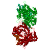
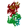
- PDB-2zlf: The Structural Basis for Peptidomimetic Inhibition of Eukaryotic ... -
+ Open data

- Basic information
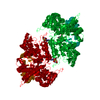
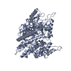
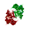
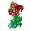
| Entry | Database: PDB / ID: 2zlf | ||||||
|---|---|---|---|---|---|---|---|
| Title | The Structural Basis for Peptidomimetic Inhibition of Eukaryotic Ribonucleotide Reductase | ||||||
 Components Components |
| ||||||
 Keywords Keywords | OXIDOREDUCTASE / peptidomimetic inhibition eukaryotic ribonucleotide reductase / Allosteric enzyme / ATP-binding / Cytoplasm / DNA replication / Nucleotide-binding / Phosphoprotein | ||||||
| Function / homology |  Function and homology information Function and homology informationInterconversion of nucleotide di- and triphosphates / ribonucleoside-diphosphate reductase complex / ribonucleoside-diphosphate reductase / ribonucleoside-diphosphate reductase activity, thioredoxin disulfide as acceptor / deoxyribonucleotide biosynthetic process / nucleotide binding / ATP binding / identical protein binding / nucleus / cytoplasm Similarity search - Function | ||||||
| Biological species |  | ||||||
| Method |  X-RAY DIFFRACTION / SYNCHROTRON / MOLECULAR REPLACEMENT / Resolution: 2.59 Å X-RAY DIFFRACTION / SYNCHROTRON / MOLECULAR REPLACEMENT / Resolution: 2.59 Å | ||||||
 Authors Authors | Xu, H. / Fairman, J.W. / Wijerathna, S.R. / LaMacchia, J. / Kreischer, N.R. / Helmbrecht, E. / Cooperman, B.S. / Dealwis, C. | ||||||
 Citation Citation | Journal: J.Med.Chem. / Year: 2008 Title: The Structural Basis for Peptidomimetic Inhibition of Eukaryotic Ribonucleotide Reductase: A Conformationally Flexible Pharmacophore Authors: Xu, H. / Fairman, J.W. / Wijerathna, S.R. / Kreischer, N.R. / LaMacchia, J. / Helmbrecht, E. / Cooperman, B.S. / Dealwis, C. | ||||||
| History |
|
- Structure visualization
| Structure viewer | Molecule: MolmilJmol/JSmol |
|---|
- Downloads & links
-Download
| PDBx/mmCIF format | 2zlf.cif.gz | 148.4 KB | Display | PDBx/mmCIF format |
|---|---|---|---|---|
| PDB format | pdb2zlf.ent.gz | 114.6 KB | Display | PDB format |
| PDBx/mmJSON format | 2zlf.json.gz | Tree view | PDBx/mmJSON format | |
| Others |  Other downloads Other downloads |
-Validation report
| Arichive directory | https://data.pdbj.org/pub/pdb/validation_reports/zl/2zlfftp://data.pdbj.org/pub/pdb/validation_reports/zl/2zlf | HTTPS FTP |
|---|
-Related structure data
| Related structure data |  2zlgC  2cvxS C: citing same article ( S: Starting model for refinement |
|---|---|
| Similar structure data |


-Links
 PDBj
PDBj
- Assembly
| Deposited unit | 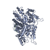
| ||||||||
|---|---|---|---|---|---|---|---|---|---|
| 1 | 
| ||||||||
| Unit cell |
|
-Components
| #1: Protein | Mass: 99672.984 Da / Num. of mol.: 1 Source method: isolated from a genetically manipulated source Source: (gene. exp.) Plasmid: PWJ751-3 / Production host:  References: UniProt: P21524, ribonucleoside-diphosphate reductase |
|---|---|
| #2: Protein/peptide | Mass: 827.878 Da / Num. of mol.: 1 / Source method: obtained synthetically |
| #3: Water | ChemComp-HOH /  Mass: 18.015 Da / Num. of mol.: 72 / Source method: isolated from a natural source / Formula: H2O Mass: 18.015 Da / Num. of mol.: 72 / Source method: isolated from a natural source / Formula: H2O |
-Experimental details
-Experiment
| Experiment | Method: X-RAY DIFFRACTION / Number of used crystals: 1 |
|---|
- Sample preparation
| Crystal | Density Matthews: 2.01 Å3/Da / Density % sol: 38.71 % |
|---|---|
| Crystal grow | Temperature: 298 K / Method: vapor diffusion, hanging drop / pH: 7.5 Details: 100mM Hepes, 20-25% PEG 3350, 0.2M NaCl, pH 7.5, VAPOR DIFFUSION, HANGING DROP, temperature 298K |
-Data collection
| Diffraction | Mean temperature: 100 K |
|---|---|
| Diffraction source | Source: SYNCHROTRON / Site: APS  / Beamline: 14-BM-C / Wavelength: 0.9002 Å / Beamline: 14-BM-C / Wavelength: 0.9002 Å |
| Detector | Type: ADSC QUANTUM 315 / Detector: CCD / Date: Mar 1, 2007 / Details: mirrors |
| Radiation | Protocol: SINGLE WAVELENGTH / Monochromatic (M) / Laue (L): M / Scattering type: x-ray |
| Radiation wavelength | Wavelength: 0.9002 Å / Relative weight: 1 |
| Reflection | Resolution: 2.59→50 Å / Num. obs: 24929 / % possible obs: 96.5 % / Observed criterion σ(I): 2 / Redundancy: 6.6 % / Rsym value: 0.081 / Net I/σ(I): 21.5 |
| Reflection shell | Resolution: 2.589→2.656 Å / Redundancy: 5.8 % / Mean I/σ(I) obs: 2.1 / Rsym value: 0.459 / % possible all: 82.4 |
- Processing
| Software |
| ||||||||||||||||||||||||||||||||||||||||||||||||||||||||||||||||||||||||||||||||||||||||||
|---|---|---|---|---|---|---|---|---|---|---|---|---|---|---|---|---|---|---|---|---|---|---|---|---|---|---|---|---|---|---|---|---|---|---|---|---|---|---|---|---|---|---|---|---|---|---|---|---|---|---|---|---|---|---|---|---|---|---|---|---|---|---|---|---|---|---|---|---|---|---|---|---|---|---|---|---|---|---|---|---|---|---|---|---|---|---|---|---|---|---|---|
| Refinement | Method to determine structure: MOLECULAR REPLACEMENT Starting model: PDB ENTRY 2CVX Resolution: 2.59→49.75 Å / Cor.coef. Fo:Fc: 0.953 / Cor.coef. Fo:Fc free: 0.92 / Cross valid method: THROUGHOUT / ESU R: 0.868 / ESU R Free: 0.322 / Stereochemistry target values: MAXIMUM LIKELIHOOD / Details: HYDROGENS HAVE BEEN ADDED IN THE RIDING POSITIONS
| ||||||||||||||||||||||||||||||||||||||||||||||||||||||||||||||||||||||||||||||||||||||||||
| Solvent computation | Ion probe radii: 0.8 Å / Shrinkage radii: 0.8 Å / VDW probe radii: 1.2 Å / Solvent model: MASK | ||||||||||||||||||||||||||||||||||||||||||||||||||||||||||||||||||||||||||||||||||||||||||
| Displacement parameters | Biso mean: 36.936 Å2
| ||||||||||||||||||||||||||||||||||||||||||||||||||||||||||||||||||||||||||||||||||||||||||
| Refinement step | Cycle: LAST / Resolution: 2.59→49.75 Å
| ||||||||||||||||||||||||||||||||||||||||||||||||||||||||||||||||||||||||||||||||||||||||||
| Refine LS restraints |
| ||||||||||||||||||||||||||||||||||||||||||||||||||||||||||||||||||||||||||||||||||||||||||
| LS refinement shell | Resolution: 2.589→2.656 Å / Total num. of bins used: 20
|
