 Movie
Movie Controller

[English] 日本語
 Yorodumi
- PDB-6mmq: Carbon regulatory PII-like protein SbtB from Cyanobium sp. 7001 b... -
+ Open data

- Basic information
| Entry | Database: PDB / ID: 6mmq | ||||||
|---|---|---|---|---|---|---|---|
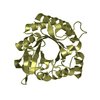
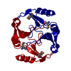
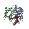
| Title | Carbon regulatory PII-like protein SbtB from Cyanobium sp. 7001 bound to cAMP | ||||||
 Components Components | Carbon regulatory PII-like protein SbtB | ||||||
 Keywords Keywords | SIGNALING PROTEIN / PII-like protein / SbtB / regulatory protein | ||||||
| Function / homology |  Function and homology information Function and homology informationregulation of nitrogen utilization / enzyme regulator activity / ATP binding / metal ion binding Similarity search - Function | ||||||
| Biological species |  Cyanobium sp. PCC 7001 (bacteria) Cyanobium sp. PCC 7001 (bacteria) | ||||||
| Method |  X-RAY DIFFRACTION / SYNCHROTRON / MOLECULAR REPLACEMENT / Resolution: 2.02 Å X-RAY DIFFRACTION / SYNCHROTRON / MOLECULAR REPLACEMENT / Resolution: 2.02 Å | ||||||
 Authors Authors | Kaczmarski, J.A. / Jackson, C. | ||||||
 Citation Citation | Journal: Biorxiv / Year: 2019 Title: Structure and function of SbtB from Cyanobium sp. 7001 Authors: Jackson, C. / Kaczmarski, J.A. / Price, D. | ||||||
| History |
|
- Structure visualization
| Structure viewer | Molecule: MolmilJmol/JSmol |
|---|
- Downloads & links
-Download
| PDBx/mmCIF format | 6mmq.cif.gz | 114.5 KB | Display | PDBx/mmCIF format |
|---|---|---|---|---|
| PDB format | pdb6mmq.ent.gz | 88.2 KB | Display | PDB format |
| PDBx/mmJSON format | 6mmq.json.gz | Tree view | PDBx/mmJSON format | |
| Others |  Other downloads Other downloads |
-Validation report
| Arichive directory | https://data.pdbj.org/pub/pdb/validation_reports/mm/6mmqftp://data.pdbj.org/pub/pdb/validation_reports/mm/6mmq | HTTPS FTP |
|---|
-Related structure data


-Links
 PDBj
PDBj


- Assembly
| Deposited unit | 
| ||||||||||
|---|---|---|---|---|---|---|---|---|---|---|---|
| 1 | 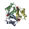
| ||||||||||
| 2 | 
| ||||||||||
| Unit cell |
|
-Components
| #1: Protein | Mass: 11484.044 Da / Num. of mol.: 6 Source method: isolated from a genetically manipulated source Source: (gene. exp.) Cyanobium sp. PCC 7001 (bacteria) / Gene: CPCC7001_1671 / Production host: #2: Chemical | ChemComp-CMP /   Mass: 329.206 Da / Num. of mol.: 6 / Source method: obtained synthetically / Formula: C10H12N5O6P Mass: 329.206 Da / Num. of mol.: 6 / Source method: obtained synthetically / Formula: C10H12N5O6P#3: Water | ChemComp-HOH / |  Mass: 18.015 Da / Num. of mol.: 34 / Source method: isolated from a natural source / Formula: H2O Mass: 18.015 Da / Num. of mol.: 34 / Source method: isolated from a natural source / Formula: H2O |
|---|
-Experimental details
-Experiment
| Experiment | Method: X-RAY DIFFRACTION / Number of used crystals: 1 |
|---|
- Sample preparation
| Crystal | Density Matthews: 2.12 Å3/Da / Density % sol: 41.91 % |
|---|---|
| Crystal grow | Temperature: 291 K / Method: vapor diffusion, hanging drop / Details: 0.2 M Potassium Acetate, 16 % PEG 3350 |
-Data collection
| Diffraction | Mean temperature: 100 K / Serial crystal experiment: N |
|---|---|
| Diffraction source | Source: SYNCHROTRON / Site: Australian Synchrotron  / Beamline: MX2 / Wavelength: 0.9537 Å / Beamline: MX2 / Wavelength: 0.9537 Å |
| Detector | Type: DECTRIS EIGER X 16M / Detector: PIXEL / Date: Aug 18, 2018 |
| Radiation | Protocol: SINGLE WAVELENGTH / Monochromatic (M) / Laue (L): M / Scattering type: x-ray |
| Radiation wavelength | Wavelength: 0.9537 Å / Relative weight: 1 |
| Reflection | Resolution: 2.02→42.48 Å / Num. obs: 35671 / % possible obs: 96.4 % / Redundancy: 2.4 % / Net I/σ(I): 6.4 |
| Reflection shell | Resolution: 2.02→2.07 Å |
- Processing
| Software |
| ||||||||||||||||||||||||||||||||||||||||||||||||||||||||||||||||||||||||||||||||||||||||||||||||||
|---|---|---|---|---|---|---|---|---|---|---|---|---|---|---|---|---|---|---|---|---|---|---|---|---|---|---|---|---|---|---|---|---|---|---|---|---|---|---|---|---|---|---|---|---|---|---|---|---|---|---|---|---|---|---|---|---|---|---|---|---|---|---|---|---|---|---|---|---|---|---|---|---|---|---|---|---|---|---|---|---|---|---|---|---|---|---|---|---|---|---|---|---|---|---|---|---|---|---|---|
| Refinement | Method to determine structure: MOLECULAR REPLACEMENT / Resolution: 2.02→42.472 Å / SU ML: 0.36 / Cross valid method: FREE R-VALUE / σ(F): 1.96 / Phase error: 37.3 / Stereochemistry target values: ML
| ||||||||||||||||||||||||||||||||||||||||||||||||||||||||||||||||||||||||||||||||||||||||||||||||||
| Solvent computation | Shrinkage radii: 0.9 Å / VDW probe radii: 1.11 Å / Solvent model: FLAT BULK SOLVENT MODEL | ||||||||||||||||||||||||||||||||||||||||||||||||||||||||||||||||||||||||||||||||||||||||||||||||||
| Refinement step | Cycle: LAST / Resolution: 2.02→42.472 Å
| ||||||||||||||||||||||||||||||||||||||||||||||||||||||||||||||||||||||||||||||||||||||||||||||||||
| Refine LS restraints |
| ||||||||||||||||||||||||||||||||||||||||||||||||||||||||||||||||||||||||||||||||||||||||||||||||||
| LS refinement shell |
|
