 Movie
Movie Controller

[English] 日本語
 Yorodumi
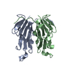
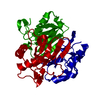
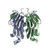
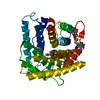
- PDB-6mm2: Carbon regulatory PII-like protein SbtB from Cyanobium sp. 7001 b... -
+ Open data

- Basic information
| Entry | Database: PDB / ID: 6mm2 | ||||||
|---|---|---|---|---|---|---|---|
| Title | Carbon regulatory PII-like protein SbtB from Cyanobium sp. 7001 bound to ATP and calcium | ||||||
 Components Components | Carbon regulatory PII-like protein SbtB | ||||||
 Keywords Keywords | SIGNALING PROTEIN / PII-like protein / SbtB / regulatory protein | ||||||
| Function / homology |  Function and homology information Function and homology informationregulation of nitrogen utilization / enzyme regulator activity / ATP binding / metal ion binding Similarity search - Function | ||||||
| Biological species |  Cyanobium sp. PCC 7001 (bacteria) Cyanobium sp. PCC 7001 (bacteria) | ||||||
| Method |  X-RAY DIFFRACTION / SYNCHROTRON / MOLECULAR REPLACEMENT / Resolution: 1.04 Å X-RAY DIFFRACTION / SYNCHROTRON / MOLECULAR REPLACEMENT / Resolution: 1.04 Å | ||||||
 Authors Authors | Kaczmarski, J.A. / Jackson, C. | ||||||
 Citation Citation | Journal: Biorxiv / Year: 2019 Title: Structure and function of SbtB from Cyanobium sp. 7001 Authors: Jackson, C. / Kaczmarski, J.A. / Price, D. | ||||||
| History |
|
- Structure visualization
| Structure viewer | Molecule: MolmilJmol/JSmol |
|---|
- Downloads & links
-Download
| PDBx/mmCIF format | 6mm2.cif.gz | 83.9 KB | Display | PDBx/mmCIF format |
|---|---|---|---|---|
| PDB format | pdb6mm2.ent.gz | 59.9 KB | Display | PDB format |
| PDBx/mmJSON format | 6mm2.json.gz | Tree view | PDBx/mmJSON format | |
| Others |  Other downloads Other downloads |
-Validation report
| Arichive directory | https://data.pdbj.org/pub/pdb/validation_reports/mm/6mm2ftp://data.pdbj.org/pub/pdb/validation_reports/mm/6mm2 | HTTPS FTP |
|---|
-Related structure data
| Related structure data |  6mmcC  6mmoC  6mmqC  6n4aC  6ntbC  5o3pS S: Starting model for refinement C: citing same article ( |
|---|---|
| Similar structure data |


-Links
 PDBj
PDBj- Assembly
| Deposited unit | 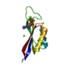
| ||||||||||||
|---|---|---|---|---|---|---|---|---|---|---|---|---|---|
| 1 | 
| ||||||||||||
| Unit cell |
| ||||||||||||
| Components on special symmetry positions |
|
-Components
| #1: Protein | Mass: 11484.044 Da / Num. of mol.: 1 Source method: isolated from a genetically manipulated source Source: (gene. exp.) Cyanobium sp. PCC 7001 (bacteria) / Gene: CPCC7001_1671 / Plasmid: pETMCSIII / Production host: |
|---|---|
| #2: Chemical | ChemComp-ATP /   Mass: 507.181 Da / Num. of mol.: 1 / Source method: obtained synthetically / Formula: C10H16N5O13P3 / Feature type: SUBJECT OF INVESTIGATION / Comment: ATP, energy-carrying molecule*YM Mass: 507.181 Da / Num. of mol.: 1 / Source method: obtained synthetically / Formula: C10H16N5O13P3 / Feature type: SUBJECT OF INVESTIGATION / Comment: ATP, energy-carrying molecule*YM |
| #3: Chemical | ChemComp-CA /   Mass: 40.078 Da / Num. of mol.: 1 / Source method: obtained synthetically / Formula: Ca / Feature type: SUBJECT OF INVESTIGATION Mass: 40.078 Da / Num. of mol.: 1 / Source method: obtained synthetically / Formula: Ca / Feature type: SUBJECT OF INVESTIGATION |
| #4: Chemical | ChemComp-CL /   Mass: 35.453 Da / Num. of mol.: 1 / Source method: obtained synthetically / Formula: Cl Mass: 35.453 Da / Num. of mol.: 1 / Source method: obtained synthetically / Formula: Cl |
| #5: Water | ChemComp-HOH /  Mass: 18.015 Da / Num. of mol.: 111 / Source method: isolated from a natural source / Formula: H2O Mass: 18.015 Da / Num. of mol.: 111 / Source method: isolated from a natural source / Formula: H2O |
| Has ligand of interest | Y |
-Experimental details
-Experiment
| Experiment | Method: X-RAY DIFFRACTION / Number of used crystals: 1 |
|---|
- Sample preparation
| Crystal | Density Matthews: 2.29 Å3/Da / Density % sol: 46.29 % |
|---|---|
| Crystal grow | Temperature: 293 K / Method: vapor diffusion, sitting drop / pH: 4.6 Details: 30 % 2-methyl-2,4-pentanediol 0.02 M calcium chloride 0.1 M sodium acetate-acetic acid pH 4.6 |
-Data collection
| Diffraction | Mean temperature: 100 K / Serial crystal experiment: N |
|---|---|
| Diffraction source | Source: SYNCHROTRON / Site: Australian Synchrotron  / Beamline: MX2 / Wavelength: 0.9537 Å / Beamline: MX2 / Wavelength: 0.9537 Å |
| Detector | Type: DECTRIS EIGER X 16M / Detector: PIXEL / Date: Jun 12, 2018 |
| Radiation | Protocol: SINGLE WAVELENGTH / Monochromatic (M) / Laue (L): M / Scattering type: x-ray |
| Radiation wavelength | Wavelength: 0.9537 Å / Relative weight: 1 |
| Reflection | Resolution: 1.04→16.045 Å / Num. obs: 44144 / % possible obs: 95.3 % / Redundancy: 9 % / CC1/2: 0.999 / Rmerge(I) obs: 0.039 / Rpim(I) all: 0.02 / Rrim(I) all: 0.043 / Χ2: 0.87 / Net I/av σ(I): 21.8 / Net I/σ(I): 0.999 |
| Reflection shell | Resolution: 1.04→1.06 Å / Redundancy: 2.2 % / Rmerge(I) obs: 1.337 / Num. unique obs: 2573 / CC1/2: 0.405 / % possible all: 50.9 |
- Processing
| Software |
| |||||||||||||||||||||
|---|---|---|---|---|---|---|---|---|---|---|---|---|---|---|---|---|---|---|---|---|---|---|
| Refinement | Method to determine structure: MOLECULAR REPLACEMENT Starting model: 5O3P Resolution: 1.04→16.04 Å / Cross valid method: FREE R-VALUE
| |||||||||||||||||||||
| Refinement step | Cycle: LAST / Resolution: 1.04→16.04 Å
| |||||||||||||||||||||
| LS refinement shell | Resolution: 1.04→1.076 Å
|
