- PDB-6mew: RFXANK ankyrin repeats in complex with a RFX7 peptide -
+
Open data
ID or keywords:
Loading...
-
Basic information
Entry
Database: PDB / ID: 6mew
Title
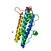
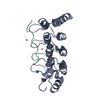
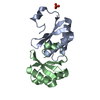
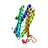
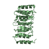
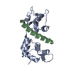
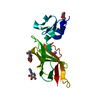
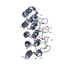
RFXANK ankyrin repeats in complex with a RFX7 peptide
Components
DNA-binding protein RFXANK
RFX7 peptide
Keywords
DNA BINDING PROTEIN / structural genomics / ankyrin repeats / Structural Genomics Consortium / SGC
Function / homology
Function and homology information
positive regulation of MHC class II biosynthetic process / RNA polymerase II core promoter sequence-specific DNA binding / RNA polymerase II transcription regulator complex / histone deacetylase binding / sequence-specific double-stranded DNA binding / DNA-binding transcription factor activity, RNA polymerase II-specific / RNA polymerase II cis-regulatory region sequence-specific DNA binding / regulation of transcription by RNA polymerase II / chromatin / DNA-templated transcription ...positive regulation of MHC class II biosynthetic process / RNA polymerase II core promoter sequence-specific DNA binding / RNA polymerase II transcription regulator complex / histone deacetylase binding / sequence-specific double-stranded DNA binding / DNA-binding transcription factor activity, RNA polymerase II-specific / RNA polymerase II cis-regulatory region sequence-specific DNA binding / regulation of transcription by RNA polymerase II / chromatin / DNA-templated transcription / positive regulation of transcription by RNA polymerase II / DNA binding / nucleoplasm / nucleus / cytoplasm Similarity search - Function
DNA-bindingproteinRFXANK / Ankyrin repeat family A protein 1 / Regulatory factor X subunit B / RFX-B / Regulatory factor X- ...Ankyrin repeat family A protein 1 / Regulatory factor X subunit B / RFX-B / Regulatory factor X-associated ankyrin-containing protein
Mass: 18926.301 Da / Num. of mol.: 2 Source method: isolated from a genetically manipulated source Source: (gene. exp.) Homo sapiens (human) / Gene: RFXANK, ANKRA1, RFXB / Plasmid: pET28-MHL / Production host: Escherichia coli (E. coli) / Strain (production host): BL21-V2R-pRARE2 / References: UniProt: O14593
#2: Protein/peptide
RFX7peptide
Mass: 2000.364 Da / Num. of mol.: 2 / Source method: obtained synthetically / Details: synthetic peptide / Source: (synth.) Homo sapiens (human) / References: UniProt: Q2KHR2*PLUS
Resolution: 1.78→34.7 Å / Cor.coef. Fo:Fc: 0.96 / Cor.coef. Fo:Fc free: 0.941 / SU B: 2.786 / SU ML: 0.087 / Cross valid method: THROUGHOUT / σ(F): 0 / ESU R: 0.123 / ESU R Free: 0.121 / Stereochemistry target values: MAXIMUM LIKELIHOOD Details: the structure of an isomorphous crystal was solved by molecular replacement, where ARP/WARP was subsequently used for phase improvement and automated model bulding. COOT was used for ...Details: the structure of an isomorphous crystal was solved by molecular replacement, where ARP/WARP was subsequently used for phase improvement and automated model bulding. COOT was used for interactive model building. Model geometry was evaluated with MOLPROBITY.
Rfactor
Num. reflection
% reflection
Selection details
Rfree
0.2187
1957
5.6 %
thin shells (sftools)
Rwork
0.1759
-
-
-
obs
0.1782
33205
98.69 %
-
Solvent computation
Ion probe radii: 0.8 Å / Shrinkage radii: 0.8 Å / VDW probe radii: 1.2 Å / Solvent model: MASK
In the structure databanks used in Yorodumi, some data are registered as the other names, "COVID-19 virus" and "2019-nCoV". Here are the details of the virus and the list of structure data.
Jan 31, 2019. EMDB accession codes are about to change! (news from PDBe EMDB page)
EMDB accession codes are about to change! (news from PDBe EMDB page)
The allocation of 4 digits for EMDB accession codes will soon come to an end. Whilst these codes will remain in use, new EMDB accession codes will include an additional digit and will expand incrementally as the available range of codes is exhausted. The current 4-digit format prefixed with “EMD-” (i.e. EMD-XXXX) will advance to a 5-digit format (i.e. EMD-XXXXX), and so on. It is currently estimated that the 4-digit codes will be depleted around Spring 2019, at which point the 5-digit format will come into force.
The EM Navigator/Yorodumi systems omit the EMD- prefix.
Related info.:Q: What is EMD? / ID/Accession-code notation in Yorodumi/EM Navigator
Yorodumi is a browser for structure data from EMDB, PDB, SASBDB, etc.
This page is also the successor to EM Navigator detail page, and also detail information page/front-end page for Omokage search.
The word "yorodu" (or yorozu) is an old Japanese word meaning "ten thousand". "mi" (miru) is to see.
Related info.:EMDB / PDB / SASBDB / Comparison of 3 databanks / Yorodumi Search / Aug 31, 2016. New EM Navigator & Yorodumi / Yorodumi Papers / Jmol/JSmol / Function and homology information / Changes in new EM Navigator and Yorodumi
 Movie
Movie Controller


 Open data
 Basic information
 Components
 Keywords
 Function and homology information
 Homo sapiens (human)
 X-RAY DIFFRACTION /
  Authors
 Citation
 Structure visualization
 Downloads & links
 Other downloads
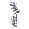


 PDBj
PDBj

 Assembly
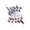


 Num. of mol.: 53 / Source method: obtained synthetically
 Mass: 18.015 Da / Num. of mol.: 246 / Source method: isolated from a natural source / Formula: H2O
 Sample preparation
 Processing