 Movie
Movie Controller

[English] 日本語
 Yorodumi
- PDB-6m3x: Cryo-EM structure of sulfur oxygenase reductase from Sulfurisphae... -
+ Open data

- Basic information
| Entry | Database: PDB / ID: 6m3x | |||||||||
|---|---|---|---|---|---|---|---|---|---|---|
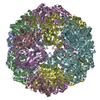
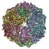
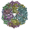
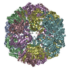
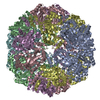
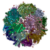
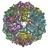
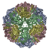
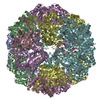
| Title | Cryo-EM structure of sulfur oxygenase reductase from Sulfurisphaera tokodaii | |||||||||
 Components Components | Sulfur oxygenase/reductase | |||||||||
 Keywords Keywords | OXIDOREDUCTASE / spherical homo 24-mer | |||||||||
| Function / homology | sulfur oxygenase/reductase / sulfur oxygenase/reductase activity / Sulphur oxygenase reductase / Sulphur oxygenase reductase / Dimeric alpha-beta barrel / metal ion binding / : / Sulfur oxygenase/reductase Function and homology information Function and homology information | |||||||||
| Biological species |   Sulfurisphaera tokodaii (archaea) Sulfurisphaera tokodaii (archaea) | |||||||||
| Method | ELECTRON MICROSCOPY / single particle reconstruction / cryo EM / Resolution: 2.24 Å | |||||||||
 Authors Authors | Sato, Y. / Adachi, N. / Moriya, T. / Arakawa, T. / Kawasaki, M. / Yamada, C. / Senda, T. / Fushinobu, S. | |||||||||
| Funding support |  Japan, 2items Japan, 2items
| |||||||||
 Citation Citation | Journal: J Struct Biol X / Year: 2020 Title: Crystallographic and cryogenic electron microscopic structures and enzymatic characterization of sulfur oxygenase reductase from . Authors: Yuta Sato / Takashi Yabuki / Naruhiko Adachi / Toshio Moriya / Takatoshi Arakawa / Masato Kawasaki / Chihaya Yamada / Toshiya Senda / Shinya Fushinobu / Takayoshi Wakagi / Abstract: Sulfur oxygenase reductases (SORs) are present in thermophilic and mesophilic archaea and bacteria, and catalyze oxygen-dependent oxygenation and disproportionation of elemental sulfur. SOR has a ...Sulfur oxygenase reductases (SORs) are present in thermophilic and mesophilic archaea and bacteria, and catalyze oxygen-dependent oxygenation and disproportionation of elemental sulfur. SOR has a hollow, spherical homo-24-mer structure and reactions take place at active sites inside the chamber. The crystal structures of SORs from species have been reported. However, the states of the active site components (mononuclear iron and cysteines) and the entry and exit paths of the substrate and products are still in dispute. Here, we report the biochemical and structural characterizations of SORs from the thermoacidophilic archaeon (StSOR) and present high-resolution structures determined by X-ray crystallography and cryogenic electron microscopy (cryo-EM). The crystal structure of StSOR was determined at 1.73 Å resolution. At the catalytic center, iron is ligated to His86, His90, Glu114, and two water molecules. Three conserved cysteines in the cavity are located 9.5-13 Å from the iron and were observed as free thiol forms. A mutational analysis indicated that the iron and one of the cysteines (Cys31) were essential for both activities. The cryo-EM structure was determined at 2.24 Å resolution using an instrument operating at 200 kV. The two structures determined by different methodologies showed similar main chain traces, but the maps exhibited different features at catalytically important components. A possible role of StSOR in the sulfur metabolism of (an obligate aerobe) is discussed based on this study. Given the high resolution achieved in this study, StSOR was shown to be a good benchmark sample for cryo-EM. | |||||||||
| History |
|
- Structure visualization
| Movie |
Movie viewer |
|---|---|
| Structure viewer | Molecule: MolmilJmol/JSmol |
- Downloads & links
-Download
| PDBx/mmCIF format | 6m3x.cif.gz | 1.3 MB | Display | PDBx/mmCIF format |
|---|---|---|---|---|
| PDB format | pdb6m3x.ent.gz | 1.1 MB | Display | PDB format |
| PDBx/mmJSON format | 6m3x.json.gz | Tree view | PDBx/mmJSON format | |
| Others |  Other downloads Other downloads |
-Validation report
| Arichive directory | https://data.pdbj.org/pub/pdb/validation_reports/m3/6m3xftp://data.pdbj.org/pub/pdb/validation_reports/m3/6m3x | HTTPS FTP |
|---|
-Related structure data
| Related structure data |  30073MC  6m35C C: citing same article ( M: map data used to model this data |
|---|---|
| Similar structure data | |
| EM raw data | EMPIAR-10546 (Title: 2.05 angstrom resolution structure determination of sulfur oxygenase reductase using 200kV cryo-EM Data size: 3.9 TB Data #1: 2.05 angstrom resolution structure determination of sulfur oxygenase reductase using 200kV cryo-EM [micrographs - multiframe]) |


-Links
 PDBj
PDBj- Assembly
| Deposited unit | 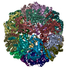
|
|---|---|
| 1 |
|
-Components
| #1: Protein | Mass: 35763.344 Da / Num. of mol.: 24 Source method: isolated from a genetically manipulated source Source: (gene. exp.) Sulfurisphaera tokodaii (strain DSM 16993 / JCM 10545 / NBRC 100140 / 7) (archaea)Strain: DSM 16993 / JCM 10545 / NBRC 100140 / 7 / Gene: sor, ST1127, STK_11270 / Plasmid: pET-17b / Production host:  #2: Chemical | ChemComp-FE /   Mass: 55.845 Da / Num. of mol.: 24 / Source method: obtained synthetically / Formula: Fe / Feature type: SUBJECT OF INVESTIGATION Mass: 55.845 Da / Num. of mol.: 24 / Source method: obtained synthetically / Formula: Fe / Feature type: SUBJECT OF INVESTIGATION#3: Water | ChemComp-HOH / |  Mass: 18.015 Da / Num. of mol.: 2243 / Source method: isolated from a natural source / Formula: H2O Mass: 18.015 Da / Num. of mol.: 2243 / Source method: isolated from a natural source / Formula: H2OHas ligand of interest | Y | |
|---|
-Experimental details
-Experiment
| Experiment | Method: ELECTRON MICROSCOPY |
|---|---|
| EM experiment | Aggregation state: PARTICLE / 3D reconstruction method: single particle reconstruction |
- Sample preparation
| Component | Name: sulfur oxygenase reductase from Sulfurisphaera tokodaii Type: ORGANELLE OR CELLULAR COMPONENT / Details: spherical homo 24-mer / Entity ID: #1 / Source: RECOMBINANT | |||||||||||||||
|---|---|---|---|---|---|---|---|---|---|---|---|---|---|---|---|---|
| Molecular weight |
| |||||||||||||||
| Source (natural) | Organism: Sulfurisphaera tokodaii (archaea) / Strain: strain 7 | |||||||||||||||
| Source (recombinant) | Organism: | |||||||||||||||
| Buffer solution | pH: 8 | |||||||||||||||
| Buffer component |
| |||||||||||||||
| Specimen | Conc.: 10 mg/ml / Embedding applied: NO / Shadowing applied: NO / Staining applied: NO / Vitrification applied: YES / Details: This sample was mono-disperse. | |||||||||||||||
| Specimen support | Details: The grid was washed by acetone prior to use. / Grid material: COPPER / Grid mesh size: 300 divisions/in. / Grid type: Quantifoil R1.2/1.3 | |||||||||||||||
| Vitrification | Instrument: FEI VITROBOT MARK IV / Cryogen name: ETHANE / Humidity: 100 % / Chamber temperature: 291 K / Details: Blotting time was 5 seconds (blot force 20) |
- Electron microscopy imaging
| Experimental equipment |  Model: Talos Arctica / Image courtesy: FEI Company |
|---|---|
| Microscopy | Model: FEI TALOS ARCTICA |
| Electron gun | Electron source:  FIELD EMISSION GUN / Accelerating voltage: 200 kV / Illumination mode: FLOOD BEAM FIELD EMISSION GUN / Accelerating voltage: 200 kV / Illumination mode: FLOOD BEAM |
| Electron lens | Mode: BRIGHT FIELD / Nominal magnification: 150000 X / Nominal defocus max: 1500 nm / Nominal defocus min: 300 nm / Cs: 2.7 mm / C2 aperture diameter: 50 µm |
| Specimen holder | Cryogen: NITROGEN |
| Image recording | Average exposure time: 50.89 sec. / Electron dose: 50 e/Å2 / Detector mode: COUNTING / Film or detector model: FEI FALCON III (4k x 4k) / Num. of grids imaged: 1 / Num. of real images: 2558 |
- Processing
| EM software |
| ||||||||||||||||||||||||||||||||||||||||||||
|---|---|---|---|---|---|---|---|---|---|---|---|---|---|---|---|---|---|---|---|---|---|---|---|---|---|---|---|---|---|---|---|---|---|---|---|---|---|---|---|---|---|---|---|---|---|
| CTF correction | Type: PHASE FLIPPING AND AMPLITUDE CORRECTION | ||||||||||||||||||||||||||||||||||||||||||||
| Particle selection | Num. of particles selected: 305182 | ||||||||||||||||||||||||||||||||||||||||||||
| Symmetry | Point symmetry: O (octahedral) | ||||||||||||||||||||||||||||||||||||||||||||
| 3D reconstruction | Resolution: 2.24 Å / Resolution method: FSC 0.143 CUT-OFF / Num. of particles: 85621 / Symmetry type: POINT | ||||||||||||||||||||||||||||||||||||||||||||
| Atomic model building | B value: 20.65 / Protocol: AB INITIO MODEL / Space: REAL / Target criteria: CCmask |
