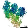
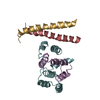
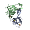
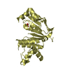
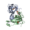
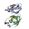
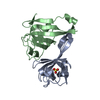
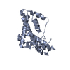
Journal: Cell Rep / Year: 2020 Title: MZT Proteins Form Multi-Faceted Structural Modules in the γ-Tubulin Ring Complex. Authors: Michal Wieczorek / Tzu-Lun Huang / Linas Urnavicius / Kuo-Chiang Hsia / Tarun M Kapoor / Abstract: Microtubule organization depends on the γ-tubulin ring complex (γ-TuRC), a ∼2.3-MDa nucleation factor comprising an asymmetric assembly of γ-tubulin and GCP2-GCP6. However, it is currently ...Microtubule organization depends on the γ-tubulin ring complex (γ-TuRC), a ∼2.3-MDa nucleation factor comprising an asymmetric assembly of γ-tubulin and GCP2-GCP6. However, it is currently unclear how the γ-TuRC-associated microproteins MZT1 and MZT2 contribute to the structure and regulation of the holocomplex. Here, we report cryo-EM structures of MZT1 and MZT2 in the context of the native human γ-TuRC. MZT1 forms two subcomplexes with the N-terminal α-helical domains of GCP3 or GCP6 (GCP-NHDs) within the γ-TuRC "lumenal bridge." We determine the X-ray structure of recombinant MZT1/GCP6-NHD and find it is similar to that within the native γ-TuRC. We identify two additional MZT/GCP-NHD-like subcomplexes, one of which is located on the outer face of the γ-TuRC and comprises MZT2 and GCP2-NHD in complex with a centrosomin motif 1 (CM1)-containing peptide. Our data reveal how MZT1 and MZT2 establish multi-faceted, structurally mimetic "modules" that can expand structural and regulatory interfaces in the γ-TuRC.
In the structure databanks used in Yorodumi, some data are registered as the other names, "COVID-19 virus" and "2019-nCoV". Here are the details of the virus and the list of structure data.
Jan 31, 2019. EMDB accession codes are about to change! (news from PDBe EMDB page)
EMDB accession codes are about to change! (news from PDBe EMDB page)
The allocation of 4 digits for EMDB accession codes will soon come to an end. Whilst these codes will remain in use, new EMDB accession codes will include an additional digit and will expand incrementally as the available range of codes is exhausted. The current 4-digit format prefixed with “EMD-” (i.e. EMD-XXXX) will advance to a 5-digit format (i.e. EMD-XXXXX), and so on. It is currently estimated that the 4-digit codes will be depleted around Spring 2019, at which point the 5-digit format will come into force.
The EM Navigator/Yorodumi systems omit the EMD- prefix.
Related info.:Q: What is EMD? / ID/Accession-code notation in Yorodumi/EM Navigator
Yorodumi is a browser for structure data from EMDB, PDB, SASBDB, etc.
This page is also the successor to EM Navigator detail page, and also detail information page/front-end page for Omokage search.
The word "yorodu" (or yorozu) is an old Japanese word meaning "ten thousand". "mi" (miru) is to see.
Related info.:EMDB / PDB / SASBDB / Comparison of 3 databanks / Yorodumi Search / Aug 31, 2016. New EM Navigator & Yorodumi / Yorodumi Papers / Jmol/JSmol / Function and homology information / Changes in new EM Navigator and Yorodumi
 Movie
Movie Controller


 Open data
 Basic information
 Components
 Keywords
 Function and homology information
 Homo sapiens (human)
 X-RAY DIFFRACTION /
  Authors
 Taiwan, 1items
  Citation
 Structure visualization
 Downloads & links
 Other downloads
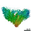


 PDBj
PDBj
 Assembly

 Sample preparation
 Processing