 Movie
Movie Controller

[English] 日本語
 Yorodumi
- PDB-6x0v: Structure of MZT2/GCP-NHD and CDK5Rap2 at position 13 of the gamm... -
+ Open data

- Basic information
| Entry | Database: PDB / ID: 6x0v | |||||||||||||||
|---|---|---|---|---|---|---|---|---|---|---|---|---|---|---|---|---|
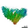
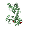
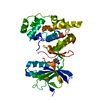
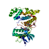
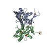
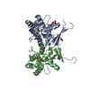
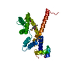
| Title | Structure of MZT2/GCP-NHD and CDK5Rap2 at position 13 of the gamma-TuRC | |||||||||||||||
 Components Components |
| |||||||||||||||
 Keywords Keywords | STRUCTURAL PROTEIN / gamma-TuRC / MZT2 / GCP / CDK5Rap2 | |||||||||||||||
| Function / homology |  Function and homology information Function and homology informationnegative regulation of centriole replication / regulation of mitotic cell cycle spindle assembly checkpoint / microtubule organizing center organization / gamma-tubulin complex / gamma-tubulin ring complex / microtubule plus-end / microtubule nucleation / microtubule bundle formation / gamma-tubulin binding / regulation of neuron differentiation ...negative regulation of centriole replication / regulation of mitotic cell cycle spindle assembly checkpoint / microtubule organizing center organization / gamma-tubulin complex / gamma-tubulin ring complex / microtubule plus-end / microtubule nucleation / microtubule bundle formation / gamma-tubulin binding / regulation of neuron differentiation / centrosome cycle / negative regulation of neuron differentiation / mitotic spindle pole / establishment of mitotic spindle orientation / pericentriolar material / centriole replication / microtubule organizing center / positive regulation of microtubule polymerization / cytoplasmic microtubule / spindle assembly / cytoplasmic microtubule organization / Loss of Nlp from mitotic centrosomes / Loss of proteins required for interphase microtubule organization from the centrosome / Recruitment of mitotic centrosome proteins and complexes / neurogenesis / Recruitment of NuMA to mitotic centrosomes / Anchoring of the basal body to the plasma membrane / AURKA Activation by TPX2 / chromosome segregation / sperm principal piece / tubulin binding / brain development / meiotic cell cycle / microtubule cytoskeleton organization / neuron migration / spindle / spindle pole / cell junction / Regulation of PLK1 Activity at G2/M Transition / mitotic cell cycle / sperm midpiece / protein-containing complex assembly / microtubule binding / microtubule / cytoskeleton / calmodulin binding / ciliary basal body / transcription cis-regulatory region binding / centrosome / protein kinase binding / positive regulation of DNA-templated transcription / protein-containing complex binding / perinuclear region of cytoplasm / Golgi apparatus / extracellular exosome / nucleoplasm / membrane / cytosol / cytoplasm Similarity search - Function | |||||||||||||||
| Biological species |  Homo sapiens (human) Homo sapiens (human) | |||||||||||||||
| Method | ELECTRON MICROSCOPY / single particle reconstruction / cryo EM / Resolution: 4.5 Å | |||||||||||||||
 Authors Authors | Wieczorek, M. / Huang, T.-L. / Urnavicius, L. / Hsia, K.-C. / Kapoor, T.M. | |||||||||||||||
| Funding support |  United States, 4items United States, 4items
| |||||||||||||||
 Citation Citation | Journal: Cell Rep / Year: 2020 Title: MZT Proteins Form Multi-Faceted Structural Modules in the γ-Tubulin Ring Complex. Authors: Michal Wieczorek / Tzu-Lun Huang / Linas Urnavicius / Kuo-Chiang Hsia / Tarun M Kapoor /  Abstract: Microtubule organization depends on the γ-tubulin ring complex (γ-TuRC), a ∼2.3-MDa nucleation factor comprising an asymmetric assembly of γ-tubulin and GCP2-GCP6. However, it is currently ...Microtubule organization depends on the γ-tubulin ring complex (γ-TuRC), a ∼2.3-MDa nucleation factor comprising an asymmetric assembly of γ-tubulin and GCP2-GCP6. However, it is currently unclear how the γ-TuRC-associated microproteins MZT1 and MZT2 contribute to the structure and regulation of the holocomplex. Here, we report cryo-EM structures of MZT1 and MZT2 in the context of the native human γ-TuRC. MZT1 forms two subcomplexes with the N-terminal α-helical domains of GCP3 or GCP6 (GCP-NHDs) within the γ-TuRC "lumenal bridge." We determine the X-ray structure of recombinant MZT1/GCP6-NHD and find it is similar to that within the native γ-TuRC. We identify two additional MZT/GCP-NHD-like subcomplexes, one of which is located on the outer face of the γ-TuRC and comprises MZT2 and GCP2-NHD in complex with a centrosomin motif 1 (CM1)-containing peptide. Our data reveal how MZT1 and MZT2 establish multi-faceted, structurally mimetic "modules" that can expand structural and regulatory interfaces in the γ-TuRC. | |||||||||||||||
| History |
|
- Structure visualization
| Movie |
Movie viewer |
|---|---|
| Structure viewer | Molecule: MolmilJmol/JSmol |
- Downloads & links
-Download
| PDBx/mmCIF format | 6x0v.cif.gz | 133.2 KB | Display | PDBx/mmCIF format |
|---|---|---|---|---|
| PDB format | pdb6x0v.ent.gz | 48.5 KB | Display | PDB format |
| PDBx/mmJSON format | 6x0v.json.gz | Tree view | PDBx/mmJSON format | |
| Others |  Other downloads Other downloads |
-Validation report
| Arichive directory | https://data.pdbj.org/pub/pdb/validation_reports/x0/6x0vftp://data.pdbj.org/pub/pdb/validation_reports/x0/6x0v | HTTPS FTP |
|---|
-Related structure data
| Related structure data | 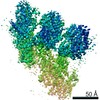 21985MC 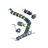 6m33C 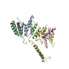 6x0uC M: map data used to model this data C: citing same article ( |
|---|---|
| Similar structure data |


-Links
 PDBj
PDBj- Assembly
| Deposited unit | 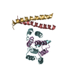
|
|---|---|
| 1 |
|
-Components
| #1: Protein | Mass: 16240.576 Da / Num. of mol.: 1 / Source method: isolated from a natural source / Source: (natural) Homo sapiens (human) / References: UniProt: Q6P582 |
|---|---|
| #2: Protein | Mass: 105765.719 Da / Num. of mol.: 1 / Source method: isolated from a natural source / Source: (natural) Homo sapiens (human) / References: UniProt: Q9BSJ2 |
| #3: Protein | Mass: 215417.359 Da / Num. of mol.: 2 / Source method: isolated from a natural source / Source: (natural) Homo sapiens (human) / References: UniProt: Q66GT8, UniProt: Q96SN8*PLUS |
-Experimental details
-Experiment
| Experiment | Method: ELECTRON MICROSCOPY |
|---|---|
| EM experiment | Aggregation state: PARTICLE / 3D reconstruction method: single particle reconstruction |
- Sample preparation
| Component | Name: Focused refinement gamma-TuRC density map surrounding positions 12-13 Type: COMPLEX / Entity ID: all / Source: NATURAL |
|---|---|
| Source (natural) | Organism: Homo sapiens (human) |
| Buffer solution | pH: 7.5 |
| Specimen | Embedding applied: NO / Shadowing applied: NO / Staining applied: NO / Vitrification applied: YES |
| Vitrification | Cryogen name: ETHANE |
- Electron microscopy imaging
| Experimental equipment |  Model: Titan Krios / Image courtesy: FEI Company |
|---|---|
| Microscopy | Model: FEI TITAN KRIOS |
| Electron gun | Electron source:  FIELD EMISSION GUN / Accelerating voltage: 300 kV / Illumination mode: OTHER FIELD EMISSION GUN / Accelerating voltage: 300 kV / Illumination mode: OTHER |
| Electron lens | Mode: BRIGHT FIELD |
| Image recording | Electron dose: 45 e/Å2 / Film or detector model: GATAN K3 (6k x 4k) |
- Processing
| Software | Name: PHENIX / Version: dev_3699: / Classification: refinement | ||||||||||||||||||||||||
|---|---|---|---|---|---|---|---|---|---|---|---|---|---|---|---|---|---|---|---|---|---|---|---|---|---|
| CTF correction | Type: PHASE FLIPPING ONLY | ||||||||||||||||||||||||
| 3D reconstruction | Resolution: 4.5 Å / Resolution method: FSC 0.143 CUT-OFF / Num. of particles: 137513 / Symmetry type: POINT | ||||||||||||||||||||||||
| Refine LS restraints |
|
