 Movie
Movie Controller


+ Open data

- Basic information
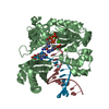
| Entry | Database: PDB / ID: 1hqv | ||||||
|---|---|---|---|---|---|---|---|
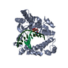
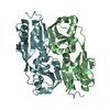
| Title | STRUCTURE OF APOPTOSIS-LINKED PROTEIN ALG-2 | ||||||
 Components Components | PROGRAMMED CELL DEATH PROTEIN 6 | ||||||
 Keywords Keywords | APOPTOSIS / penta-EF-hand protein / calcium binding protein | ||||||
| Function / homology |  Function and homology information Function and homology informationneural crest formation / vascular endothelial growth factor receptor-2 signaling pathway / neural crest cell development / COPII vesicle coat assembly / COPII vesicle coat / negative regulation of TOR signaling / positive regulation of protein monoubiquitination / negative regulation of vascular endothelial growth factor receptor signaling pathway / Cul3-RING ubiquitin ligase complex / endoplasmic reticulum exit site ...neural crest formation / vascular endothelial growth factor receptor-2 signaling pathway / neural crest cell development / COPII vesicle coat assembly / COPII vesicle coat / negative regulation of TOR signaling / positive regulation of protein monoubiquitination / negative regulation of vascular endothelial growth factor receptor signaling pathway / Cul3-RING ubiquitin ligase complex / endoplasmic reticulum exit site / endoplasmic reticulum to Golgi vesicle-mediated transport / ubiquitin-like ligase-substrate adaptor activity / protein-membrane adaptor activity / positive regulation of endothelial cell proliferation / positive regulation of endothelial cell migration / negative regulation of phosphatidylinositol 3-kinase/protein kinase B signal transduction / intracellular protein transport / response to calcium ion / positive regulation of angiogenesis / calcium-dependent protein binding / cellular response to heat / angiogenesis / cytoplasmic vesicle / protein-macromolecule adaptor activity / endosome / protein dimerization activity / positive regulation of apoptotic process / protein heterodimerization activity / calcium ion binding / apoptotic process / endoplasmic reticulum membrane / perinuclear region of cytoplasm / magnesium ion binding / endoplasmic reticulum / protein homodimerization activity / nucleoplasm / identical protein binding / nucleus / cytosol / cytoplasm Similarity search - Function | ||||||
| Biological species |  | ||||||
| Method |  X-RAY DIFFRACTION / SYNCHROTRON / MIR / Resolution: 2.3 Å X-RAY DIFFRACTION / SYNCHROTRON / MIR / Resolution: 2.3 Å | ||||||
 Authors Authors | Jia, J. / Tarabykina, S. / Hansen, C. / Berchtold, M. / Cygler, M. | ||||||
 Citation Citation | Journal: Structure / Year: 2001 Title: Structure of apoptosis-linked protein ALG-2: insights into Ca2+-induced changes in penta-EF-hand proteins. Authors: Jia, J. / Tarabykina, S. / Hansen, C. / Berchtold, M. / Cygler, M. | ||||||
| History |
|
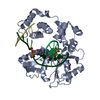
- Structure visualization
| Structure viewer | Molecule: MolmilJmol/JSmol |
|---|
- Downloads & links
-Download
| PDBx/mmCIF format | 1hqv.cif.gz | 51 KB | Display | PDBx/mmCIF format |
|---|---|---|---|---|
| PDB format | pdb1hqv.ent.gz | 36.4 KB | Display | PDB format |
| PDBx/mmJSON format | 1hqv.json.gz | Tree view | PDBx/mmJSON format | |
| Others |  Other downloads Other downloads |
-Validation report
| Arichive directory | https://data.pdbj.org/pub/pdb/validation_reports/hq/1hqvftp://data.pdbj.org/pub/pdb/validation_reports/hq/1hqv | HTTPS FTP |
|---|
-Related structure data
| Similar structure data |
|---|


-Links
 PDBj
PDBj- Assembly
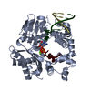
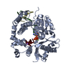
| Deposited unit | 
| ||||||||||||
|---|---|---|---|---|---|---|---|---|---|---|---|---|---|
| 1 | 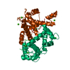
| ||||||||||||
| 2 | 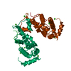
| ||||||||||||
| 3 | 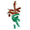
| ||||||||||||
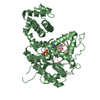
| Unit cell |
| ||||||||||||
| Components on special symmetry positions |
|
-Components
| #1: Protein | Mass: 21888.396 Da / Num. of mol.: 1 Source method: isolated from a genetically manipulated source Source: (gene. exp.)  | ||
|---|---|---|---|
| #2: Chemical | ChemComp-CA /   Mass: 40.078 Da / Num. of mol.: 4 / Source method: obtained synthetically / Formula: Ca Mass: 40.078 Da / Num. of mol.: 4 / Source method: obtained synthetically / Formula: Ca#3: Water | ChemComp-HOH / |  Mass: 18.015 Da / Num. of mol.: 51 / Source method: isolated from a natural source / Formula: H2O Mass: 18.015 Da / Num. of mol.: 51 / Source method: isolated from a natural source / Formula: H2O |
-Experimental details
-Experiment
| Experiment | Method: X-RAY DIFFRACTION / Number of used crystals: 1 |
|---|
- Sample preparation
| Crystal | Density Matthews: 2.71 Å3/Da / Density % sol: 54.66 % | ||||||||||||||||||||||||||||||
|---|---|---|---|---|---|---|---|---|---|---|---|---|---|---|---|---|---|---|---|---|---|---|---|---|---|---|---|---|---|---|---|
| Crystal grow | Temperature: 293 K / Method: vapor diffusion, hanging drop / pH: 6.5 Details: PEG 8000, Calcium Acetate, Sodium cacodylate, pH 6.5, VAPOR DIFFUSION, HANGING DROP, temperature 293K | ||||||||||||||||||||||||||||||
| Crystal grow | *PLUS | ||||||||||||||||||||||||||||||
| Components of the solutions | *PLUS
|
-Data collection
| Diffraction | Mean temperature: 100 K |
|---|---|
| Diffraction source | Source: SYNCHROTRON / Site: NSLS  / Beamline: X8C / Wavelength: 1.04453 Å / Beamline: X8C / Wavelength: 1.04453 Å |
| Detector | Type: ADSC QUANTUM 4 / Detector: CCD / Date: Sep 8, 1999 |
| Radiation | Protocol: SINGLE WAVELENGTH / Monochromatic (M) / Laue (L): M / Scattering type: x-ray |
| Radiation wavelength | Wavelength: 1.04453 Å / Relative weight: 1 |
| Reflection | Resolution: 2.3→30 Å / Num. all: 11288 / Num. obs: 10911 / % possible obs: 96.8 % / Redundancy: 6.4 % / Biso Wilson estimate: 50.16 Å2 / Rmerge(I) obs: 0.061 / Rsym value: 0.063 / Net I/σ(I): 23.7 |
| Reflection shell | Resolution: 2.3→2.35 Å / Rmerge(I) obs: 0.213 / Num. unique all: 699 / Rsym value: 0.233 / % possible all: 96 |
| Reflection | *PLUS Num. measured all: 69890 |
| Reflection shell | *PLUS % possible obs: 96 % |
- Processing
| Software |
| |||||||||||||||||||||||||
|---|---|---|---|---|---|---|---|---|---|---|---|---|---|---|---|---|---|---|---|---|---|---|---|---|---|---|
| Refinement | Method to determine structure: MIR / Resolution: 2.3→30 Å / Stereochemistry target values: Engh & Huber
| |||||||||||||||||||||||||
| Refinement step | Cycle: LAST / Resolution: 2.3→30 Å
| |||||||||||||||||||||||||
| Refine LS restraints |
| |||||||||||||||||||||||||
| Software | *PLUS Name: CNS / Version: 1 / Classification: refinement | |||||||||||||||||||||||||
| Refinement | *PLUS | |||||||||||||||||||||||||
| Solvent computation | *PLUS | |||||||||||||||||||||||||
| Displacement parameters | *PLUS Biso mean: 44.7 Å2 | |||||||||||||||||||||||||
| Refine LS restraints | *PLUS Type: c_angle_deg / Dev ideal: 1.22 |
