 Movie
Movie Controller


+ Open data

- Basic information
| Entry | Database: PDB / ID: 1aw8 | |||||||||
|---|---|---|---|---|---|---|---|---|---|---|
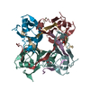
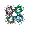
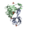
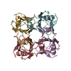
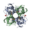
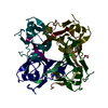
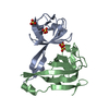
| Title | PYRUVOYL DEPENDENT ASPARTATE DECARBOXYLASE | |||||||||
 Components Components | (L-ASPARTATE-ALPHA-DECARBOXYLASE) x 2 | |||||||||
 Keywords Keywords | DECARBOXYLASE / PANTOTHENATE PATHWAY / LYASE / PROTEIN SELF-PROCESSING | |||||||||
| Function / homology |  Function and homology information Function and homology informationalanine biosynthetic process / aspartate 1-decarboxylase / aspartate 1-decarboxylase activity / pantothenate biosynthetic process / protein autoprocessing / cytosol Similarity search - Function | |||||||||
| Biological species |  | |||||||||
| Method |  X-RAY DIFFRACTION / SYNCHROTRON / MIR / Resolution: 2.2 Å X-RAY DIFFRACTION / SYNCHROTRON / MIR / Resolution: 2.2 Å | |||||||||
 Authors Authors | Albert, A. / Dhanaraj, V. / Genschel, U. / Khan, G. / Ramjee, M.K. / Pulido, R. / Sybanda, B.L. / von Delf, F. / Witty, M. / Blundell, T.L. ...Albert, A. / Dhanaraj, V. / Genschel, U. / Khan, G. / Ramjee, M.K. / Pulido, R. / Sybanda, B.L. / von Delf, F. / Witty, M. / Blundell, T.L. / Smith, A.G. / Abell, C. | |||||||||
 Citation Citation | Journal: Nat.Struct.Biol. / Year: 1998 Title: Crystal structure of aspartate decarboxylase at 2.2 A resolution provides evidence for an ester in protein self-processing. Authors: Albert, A. / Dhanaraj, V. / Genschel, U. / Khan, G. / Ramjee, M.K. / Pulido, R. / Sibanda, B.L. / von Delft, F. / Witty, M. / Blundell, T.L. / Smith, A.G. / Abell, C. | |||||||||
| History |
|
- Structure visualization
| Structure viewer | Molecule: MolmilJmol/JSmol |
|---|
- Downloads & links
-Download
| PDBx/mmCIF format | 1aw8.cif.gz | 59.1 KB | Display | PDBx/mmCIF format |
|---|---|---|---|---|
| PDB format | pdb1aw8.ent.gz | 43.8 KB | Display | PDB format |
| PDBx/mmJSON format | 1aw8.json.gz | Tree view | PDBx/mmJSON format | |
| Others |  Other downloads Other downloads |
-Validation report
| Arichive directory | https://data.pdbj.org/pub/pdb/validation_reports/aw/1aw8ftp://data.pdbj.org/pub/pdb/validation_reports/aw/1aw8 | HTTPS FTP |
|---|
-Related structure data
| Similar structure data |
|---|


-Links
 PDBj
PDBj


- Assembly
| Deposited unit | 
| ||||||||
|---|---|---|---|---|---|---|---|---|---|
| 1 | 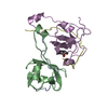
| ||||||||
| Unit cell |
| ||||||||
| Noncrystallographic symmetry (NCS) | NCS oper: (Code: given Matrix: (0.764933, 0.394656, -0.509043), Vector: |
-Components
| #1: Protein/peptide | Mass: 2841.380 Da / Num. of mol.: 2 Source method: isolated from a genetically manipulated source Source: (gene. exp.) #2: Protein | Mass: 9872.942 Da / Num. of mol.: 2 Source method: isolated from a genetically manipulated source Details: microheterogeneity at residue B25 / Source: (gene. exp.) #3: Water | ChemComp-HOH / |  Mass: 18.015 Da / Num. of mol.: 98 / Source method: isolated from a natural source / Formula: H2O Mass: 18.015 Da / Num. of mol.: 98 / Source method: isolated from a natural source / Formula: H2OHas protein modification | Y | |
|---|
-Experimental details
-Experiment
| Experiment | Method: X-RAY DIFFRACTION / Number of used crystals: 1 |
|---|
- Sample preparation
| Crystal | Density Matthews: 3.2 Å3/Da / Density % sol: 65 % | |||||||||||||||||||||||||
|---|---|---|---|---|---|---|---|---|---|---|---|---|---|---|---|---|---|---|---|---|---|---|---|---|---|---|
| Crystal grow | pH: 4.6 Details: PROTEIN WAS CRYSTALLIZED FROM 12% PEG 2000 MME, 0.1 M NA ACETATE, PH 4.6 | |||||||||||||||||||||||||
| Crystal | *PLUS | |||||||||||||||||||||||||
| Crystal grow | *PLUS pH: 7.5 / Method: vapor diffusion | |||||||||||||||||||||||||
| Components of the solutions | *PLUS
|
-Data collection
| Diffraction | Mean temperature: 293 K |
|---|---|
| Diffraction source | Source: SYNCHROTRON / Site: SRS  / Beamline: PX9.6 / Wavelength: 1.488 / Beamline: PX9.6 / Wavelength: 1.488 |
| Detector | Date: Jul 31, 1996 |
| Radiation | Monochromatic (M) / Laue (L): M / Scattering type: x-ray |
| Radiation wavelength | Wavelength: 1.488 Å / Relative weight: 1 |
| Reflection | Resolution: 2.2→23.6 Å / Num. obs: 16129 / % possible obs: 98 % / Observed criterion σ(I): 2.2 / Redundancy: 3.7 % / Rmerge(I) obs: 0.078 / Net I/σ(I): 6.4 |
| Reflection shell | Resolution: 2.2→2.26 Å / Redundancy: 3.6 % / Rmerge(I) obs: 0.191 / Mean I/σ(I) obs: 3.6 / % possible all: 80 |
| Reflection shell | *PLUS % possible obs: 90 % |
- Processing
| Software |
| |||||||||||||||||||||
|---|---|---|---|---|---|---|---|---|---|---|---|---|---|---|---|---|---|---|---|---|---|---|
| Refinement | Method to determine structure: MIR / Resolution: 2.2→8 Å / Rfactor Rfree error: 0.007 / Data cutoff high absF: 100000 / Data cutoff low absF: 0.1 / Cross valid method: THROUGHOUT / σ(F): 2.2 Details: THE ELECTRON DENSITY FOR RESIDUES 20 - 25 DIFFERS IN THE TWO PROTOMERS IN THE ASYMMETRIC UNIT. THE FIRST PROTOMER (CHAINS D AND E) SHOWS CLEAR INDICATION FOR A PYRUVOYL GROUP. IN THE SECOND ...Details: THE ELECTRON DENSITY FOR RESIDUES 20 - 25 DIFFERS IN THE TWO PROTOMERS IN THE ASYMMETRIC UNIT. THE FIRST PROTOMER (CHAINS D AND E) SHOWS CLEAR INDICATION FOR A PYRUVOYL GROUP. IN THE SECOND (CHAINS A AND B), ELECTRON DENSITY INDICATES TWO SUPERPOSED STRUCTURES OF EQUAL OCCUPANCY, ONE CORRESPONDING TO A PYRUVOYL GROUP (RESIDUE PVL) AND THE OTHER (RESIDUE SEG) CORRESPONDING TO AN ESTER INTERMEDIATE IN THE FORMATION OF THE PYRUVOYL GROUP. HET GROUP SEG LINKS CHAINS A AND B. BECAUSE OF PDB FORMAT LIMITATIONS ONLY PVL APPEARS ON SEQRES. THE ELECTRON DENSITY FOR RESIDUES 20 - 25 DIFFERS IN THE TWO PROTOMERS IN THE ASYMMETRIC UNIT. THE FIRST PROTOMER (CHAINS D AND E) SHOWS CLEAR INDICATION FOR A PYRUVOYL GROUP. IN THE SECOND (CHAINS A AND B), ELECTRON DENSITY INDICATES TWO SUPERPOSED STRUCTURES OF EQUAL OCCUPANCY, ONE CORRESPONDING TO A PYRUVOYL GROUP (RESIDUE PVL) AND THE OTHER (RESIDUE SEG) CORRESPONDING TO AN ESTER INTERMEDIATE IN THE FORMATION OF THE PYRUVOYL GROUP. HET GROUP SEG LINKS CHAINS A AND B. BECAUSE OF PDB FORMAT LIMITATIONS ONLY PVL APPEARS ON SEQRES.
| |||||||||||||||||||||
| Refinement step | Cycle: LAST / Resolution: 2.2→8 Å
| |||||||||||||||||||||
| LS refinement shell | Resolution: 2.2→2.3 Å / Rfactor Rfree error: 0.02 / Total num. of bins used: 8 /
| |||||||||||||||||||||
| Xplor file |
| |||||||||||||||||||||
| Software | *PLUS Name: X-PLOR / Version: 3.843 / Classification: refinement | |||||||||||||||||||||
| Refinement | *PLUS % reflection Rfree: 7 % / Rfactor obs: 0.2 / Rfactor Rwork: 0.2 | |||||||||||||||||||||
| Solvent computation | *PLUS | |||||||||||||||||||||
| Displacement parameters | *PLUS | |||||||||||||||||||||
| Refine LS restraints | *PLUS
|
