 Movie
Movie Controller

[English] 日本語
 Yorodumi
- PDB-6l47: Structure of the human sterol O-acyltransferase 1 in complex with... -
+ Open data

- Basic information
| Entry | Database: PDB / ID: 6l47 | ||||||||||||||||||
|---|---|---|---|---|---|---|---|---|---|---|---|---|---|---|---|---|---|---|---|
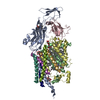
| Title | Structure of the human sterol O-acyltransferase 1 in complex with CI-976 | ||||||||||||||||||
 Components Components | Sterol O-acyltransferase 1 | ||||||||||||||||||
 Keywords Keywords | MEMBRANE PROTEIN / TRANSFERASE / SOAT / ACAT / MBOAT | ||||||||||||||||||
| Function / homology |  Function and homology information Function and homology informationsterol O-acyltransferase / sterol O-acyltransferase activity / cholesterol O-acyltransferase activity / cholesterol storage / macrophage derived foam cell differentiation / positive regulation of amyloid precursor protein biosynthetic process / very-low-density lipoprotein particle assembly / fatty-acyl-CoA binding / low-density lipoprotein particle clearance / LDL clearance ...sterol O-acyltransferase / sterol O-acyltransferase activity / cholesterol O-acyltransferase activity / cholesterol storage / macrophage derived foam cell differentiation / positive regulation of amyloid precursor protein biosynthetic process / very-low-density lipoprotein particle assembly / fatty-acyl-CoA binding / low-density lipoprotein particle clearance / LDL clearance / cholesterol efflux / cholesterol binding / cholesterol metabolic process / cholesterol homeostasis / endoplasmic reticulum membrane / endoplasmic reticulum / membrane / identical protein binding Similarity search - Function | ||||||||||||||||||
| Biological species |  Homo sapiens (human) Homo sapiens (human) | ||||||||||||||||||
| Method | ELECTRON MICROSCOPY / single particle reconstruction / cryo EM / Resolution: 3.5 Å | ||||||||||||||||||
 Authors Authors | Chen, L. / Guan, C. / Niu, Y. | ||||||||||||||||||
| Funding support |  China, 5items China, 5items
| ||||||||||||||||||
 Citation Citation | Journal: Nat Commun / Year: 2020 Title: Structural insights into the inhibition mechanism of human sterol O-acyltransferase 1 by a competitive inhibitor. Authors: Chengcheng Guan / Yange Niu / Si-Cong Chen / Yunlu Kang / Jing-Xiang Wu / Koji Nishi / Catherine C Y Chang / Ta-Yuan Chang / Tuoping Luo / Lei Chen /  Abstract: Sterol O-acyltransferase 1 (SOAT1) is an endoplasmic reticulum (ER) resident, multi-transmembrane enzyme that belongs to the membrane-bound O-acyltransferase (MBOAT) family. It catalyzes the ...Sterol O-acyltransferase 1 (SOAT1) is an endoplasmic reticulum (ER) resident, multi-transmembrane enzyme that belongs to the membrane-bound O-acyltransferase (MBOAT) family. It catalyzes the esterification of cholesterol to generate cholesteryl esters for cholesterol storage. SOAT1 is a target to treat several human diseases. However, its structure and mechanism remain elusive since its discovery. Here, we report the structure of human SOAT1 (hSOAT1) determined by cryo-EM. hSOAT1 is a tetramer consisted of a dimer of dimer. The structure of hSOAT1 dimer at 3.5 Å resolution reveals that a small molecule inhibitor CI-976 binds inside the catalytic chamber and blocks the accessibility of the active site residues H460, N421 and W420. Our results pave the way for future mechanistic study and rational drug design targeting hSOAT1 and other mammalian MBOAT family members. | ||||||||||||||||||
| History |
|
- Structure visualization
| Movie |
Movie viewer |
|---|---|
| Structure viewer | Molecule: MolmilJmol/JSmol |
- Downloads & links
-Download
| PDBx/mmCIF format | 6l47.cif.gz | 184.5 KB | Display | PDBx/mmCIF format |
|---|---|---|---|---|
| PDB format | pdb6l47.ent.gz | 142.6 KB | Display | PDB format |
| PDBx/mmJSON format | 6l47.json.gz | Tree view | PDBx/mmJSON format | |
| Others |  Other downloads Other downloads |
-Validation report
| Arichive directory | https://data.pdbj.org/pub/pdb/validation_reports/l4/6l47ftp://data.pdbj.org/pub/pdb/validation_reports/l4/6l47 | HTTPS FTP |
|---|
-Related structure data
| Related structure data |  0831MC 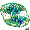 0829C 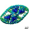 0830C  0832C  6l48C M: map data used to model this data C: citing same article ( |
|---|---|
| Similar structure data |


-Links
 PDBj
PDBj


- Assembly
| Deposited unit | 
|
|---|---|
| 1 |
|
-Components
| #1: Protein | Mass: 57294.777 Da / Num. of mol.: 2 Source method: isolated from a genetically manipulated source Source: (gene. exp.) Homo sapiens (human) / Gene: SOAT1, ACACT, ACACT1, ACAT, ACAT1, SOAT, STAT / Production host: Homo sapiens (human) / References: UniProt: P35610, sterol O-acyltransferase#2: Chemical |   Mass: 393.560 Da / Num. of mol.: 2 / Source method: obtained synthetically / Formula: C23H39NO4 / Feature type: SUBJECT OF INVESTIGATION Mass: 393.560 Da / Num. of mol.: 2 / Source method: obtained synthetically / Formula: C23H39NO4 / Feature type: SUBJECT OF INVESTIGATION#3: Chemical |   Mass: 386.654 Da / Num. of mol.: 2 / Source method: obtained synthetically / Formula: C27H46O Mass: 386.654 Da / Num. of mol.: 2 / Source method: obtained synthetically / Formula: C27H46OHas ligand of interest | Y | Has protein modification | Y | |
|---|
-Experimental details
-Experiment
| Experiment | Method: ELECTRON MICROSCOPY |
|---|---|
| EM experiment | Aggregation state: PARTICLE / 3D reconstruction method: single particle reconstruction |
- Sample preparation
| Component | Name: human sterol O-acyltransferase 1 dimer / Type: COMPLEX / Entity ID: #1 / Source: RECOMBINANT |
|---|---|
| Source (natural) | Organism: Homo sapiens (human) |
| Source (recombinant) | Organism: Homo sapiens (human) |
| Buffer solution | pH: 7.5 |
| Specimen | Embedding applied: NO / Shadowing applied: NO / Staining applied: NO / Vitrification applied: YES |
| Vitrification | Cryogen name: ETHANE |
- Electron microscopy imaging
| Experimental equipment |  Model: Titan Krios / Image courtesy: FEI Company |
|---|---|
| Microscopy | Model: FEI TITAN KRIOS |
| Electron gun | Electron source:  FIELD EMISSION GUN / Accelerating voltage: 300 kV / Illumination mode: FLOOD BEAM FIELD EMISSION GUN / Accelerating voltage: 300 kV / Illumination mode: FLOOD BEAM |
| Electron lens | Mode: BRIGHT FIELD |
| Image recording | Electron dose: 50 e/Å2 / Film or detector model: GATAN K2 QUANTUM (4k x 4k) |
- Processing
| EM software | Name: cisTEM / Category: 3D reconstruction |
|---|---|
| CTF correction | Type: NONE |
| Symmetry | Point symmetry: C2 (2 fold cyclic) |
| 3D reconstruction | Resolution: 3.5 Å / Resolution method: FSC 0.143 CUT-OFF / Num. of particles: 222018 / Details: cisTEM resolution limiting methods / Symmetry type: POINT |
