 Movie
Movie Controller

[English] 日本語
 Yorodumi
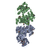
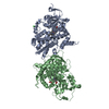
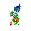
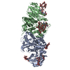
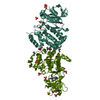
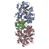
- PDB-1gqy: MURC - CRYSTAL STRUCTURE OF THE ENZYME FROM HAEMOPHILUS INFLUENZA... -
+ Open data

- Basic information
| Entry | Database: PDB / ID: 1gqy | ||||||
|---|---|---|---|---|---|---|---|
| Title | MURC - CRYSTAL STRUCTURE OF THE ENZYME FROM HAEMOPHILUS INFLUENZAE COMPLEXED WITH AMPPCP | ||||||
 Components Components | UDP-N-ACETYLMURAMATE-L-ALANINE LIGASE | ||||||
 Keywords Keywords | CELL WALL BIOSYNTHESIS / PEPTIDOGLYCAN / MUREIN / LIGASE / ATP BINDING | ||||||
| Function / homology |  Function and homology information Function and homology informationUDP-N-acetylmuramate-L-alanine ligase / UDP-N-acetylmuramate-L-alanine ligase activity / peptidoglycan biosynthetic process / cell wall organization / regulation of cell shape / cell division / ATP binding / cytoplasm Similarity search - Function | ||||||
| Biological species |  HAEMOPHILUS INFLUENZAE (bacteria) HAEMOPHILUS INFLUENZAE (bacteria) | ||||||
| Method |  X-RAY DIFFRACTION / SYNCHROTRON / MOLECULAR REPLACEMENT / Resolution: 1.8 Å X-RAY DIFFRACTION / SYNCHROTRON / MOLECULAR REPLACEMENT / Resolution: 1.8 Å | ||||||
 Authors Authors | Skarzynski, T. / Cleasby, A. / Domenici, E. / Gevi, M. / Shaw, J. | ||||||
 Citation Citation | Journal: To be Published Title: Crystal Structures of Udp-N-Acetylmuramate-L-Alanine Ligase (Murc) from Haemophilus Influenzae Authors: Skarzynski, T. / Cleasby, A. / Domenici, E. / Gevi, M. / Shaw, J. | ||||||
| History |
|
- Structure visualization
| Structure viewer | Molecule: MolmilJmol/JSmol |
|---|
- Downloads & links
-Download
| PDBx/mmCIF format | 1gqy.cif.gz | 209.8 KB | Display | PDBx/mmCIF format |
|---|---|---|---|---|
| PDB format | pdb1gqy.ent.gz | 167.2 KB | Display | PDB format |
| PDBx/mmJSON format | 1gqy.json.gz | Tree view | PDBx/mmJSON format | |
| Others |  Other downloads Other downloads |
-Validation report
| Arichive directory | https://data.pdbj.org/pub/pdb/validation_reports/gq/1gqyftp://data.pdbj.org/pub/pdb/validation_reports/gq/1gqy | HTTPS FTP |
|---|
-Related structure data
| Related structure data | 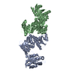 1gqqSC S: Starting model for refinement C: citing same article ( |
|---|---|
| Similar structure data |


-Links
 PDBj
PDBj


- Assembly
| Deposited unit | 
| ||||||||
|---|---|---|---|---|---|---|---|---|---|
| 1 |
| ||||||||
| Unit cell |
|
-Components
| #1: Protein | Mass: 52057.172 Da / Num. of mol.: 2 Source method: isolated from a genetically manipulated source Source: (gene. exp.) HAEMOPHILUS INFLUENZAE (bacteria) / Production host: References: UniProt: P45066, UDP-N-acetylmuramate-L-alanine ligase #2: Chemical |   Mass: 505.208 Da / Num. of mol.: 2 / Source method: obtained synthetically / Formula: C11H18N5O12P3 / Comment: AMP-PCP, energy-carrying molecule analogue*YM Mass: 505.208 Da / Num. of mol.: 2 / Source method: obtained synthetically / Formula: C11H18N5O12P3 / Comment: AMP-PCP, energy-carrying molecule analogue*YM#3: Chemical |   Mass: 24.305 Da / Num. of mol.: 2 / Source method: obtained synthetically / Formula: Mg Mass: 24.305 Da / Num. of mol.: 2 / Source method: obtained synthetically / Formula: Mg#4: Water | ChemComp-HOH / |  Mass: 18.015 Da / Num. of mol.: 925 / Source method: isolated from a natural source / Formula: H2O Mass: 18.015 Da / Num. of mol.: 925 / Source method: isolated from a natural source / Formula: H2O |
|---|
-Experimental details
-Experiment
| Experiment | Method: X-RAY DIFFRACTION / Number of used crystals: 1 |
|---|
- Sample preparation
| Crystal | Density Matthews: 2.73 Å3/Da / Density % sol: 54.89 % |
|---|---|
| Crystal grow | pH: 6.8 Details: THE AMPPCP COMPLEX WAS FORMED BY ADDING 10MM AMPPCP AND 10MM MGCL2 TO THE CONCENTRATED PROTEIN SOLUTION AT 12MG/ML. CRYSTALS WERE GROWN USING WELL SOLUTION MADE OF 20%PEG 3350 AND 200MM SODIUM FORMATE, PH 6.8. |
-Data collection
| Diffraction | Mean temperature: 100 K |
|---|---|
| Diffraction source | Source: SYNCHROTRON / Site: SRS  / Beamline: PX9.6 / Wavelength: 0.87 / Beamline: PX9.6 / Wavelength: 0.87 |
| Detector | Type: ADSC CCD / Detector: CCD / Date: Oct 7, 1999 |
| Radiation | Protocol: SINGLE WAVELENGTH / Monochromatic (M) / Laue (L): M / Scattering type: x-ray |
| Radiation wavelength | Wavelength: 0.87 Å / Relative weight: 1 |
| Reflection | Resolution: 1.8→30 Å / Num. obs: 93715 / % possible obs: 89.5 % / Redundancy: 2 % / Rmerge(I) obs: 0.069 |
| Reflection shell | Resolution: 1.8→1.86 Å / Rmerge(I) obs: 0.453 / % possible all: 47.7 |
- Processing
| Software |
| ||||||||||||||||||||
|---|---|---|---|---|---|---|---|---|---|---|---|---|---|---|---|---|---|---|---|---|---|
| Refinement | Method to determine structure: MOLECULAR REPLACEMENT Starting model: AN EARLY VERSION OF PDB ENTRY 1GQQ Resolution: 1.8→20 Å / SU B: 3.26087 / SU ML: 0.09953 / Cross valid method: THROUGHOUT / σ(F): 0 / ESU R: 0.12872 / ESU R Free: 0.12945 Details: DISORDERED SIDE CHAINS WERE MODELLED STEREOCHEMICALLY
| ||||||||||||||||||||
| Displacement parameters | Biso mean: 30.035 Å2
| ||||||||||||||||||||
| Refinement step | Cycle: LAST / Resolution: 1.8→20 Å
|
