 Movie
Movie Controller

[English] 日本語
 Yorodumi
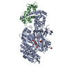
- PDB-5nmx: Crystal Structure of the pyrrolizidine alkaloid N-oxygenase from ... -
+ Open data

- Basic information
| Entry | Database: PDB / ID: 5nmx | ||||||
|---|---|---|---|---|---|---|---|
| Title | Crystal Structure of the pyrrolizidine alkaloid N-oxygenase from Zonocerus variegatus in complex with FAD and NADP+ | ||||||
 Components Components | Flavin-containing monooxygenase | ||||||
 Keywords Keywords | OXIDOREDUCTASE / pyrrolizidine alkaloid N-oxygenase / flavin-containing monooxygenase / rossmann fold / FAD / NADP / two dinucleotide binding domain flavoprotein / senecionine | ||||||
| Function / homology |  Function and homology information Function and homology informationN,N-dimethylaniline monooxygenase activity / Oxidoreductases / NADP binding / flavin adenine dinucleotide binding Similarity search - Function | ||||||
| Biological species |  Zonocerus variegatus (insect) Zonocerus variegatus (insect) | ||||||
| Method |  X-RAY DIFFRACTION / SYNCHROTRON / MOLECULAR REPLACEMENT / Resolution: 1.6 Å X-RAY DIFFRACTION / SYNCHROTRON / MOLECULAR REPLACEMENT / Resolution: 1.6 Å | ||||||
 Authors Authors | Scheidig, A. / Kubitza, C. / Faust, A. / Ober, D. | ||||||
 Citation Citation | Journal: Acta Crystallogr D Struct Biol / Year: 2018 Title: Crystal structure of pyrrolizidine alkaloid N-oxygenase from the grasshopper Zonocerus variegatus. Authors: Kubitza, C. / Faust, A. / Gutt, M. / Gath, L. / Ober, D. / Scheidig, A.J. | ||||||
| History |
|
- Structure visualization
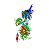
| Structure viewer | Molecule: MolmilJmol/JSmol |
|---|
- Downloads & links
-Download
| PDBx/mmCIF format | 5nmx.cif.gz | 386.3 KB | Display | PDBx/mmCIF format |
|---|---|---|---|---|
| PDB format | pdb5nmx.ent.gz | 310.3 KB | Display | PDB format |
| PDBx/mmJSON format | 5nmx.json.gz | Tree view | PDBx/mmJSON format | |
| Others |  Other downloads Other downloads |
-Validation report
| Arichive directory | https://data.pdbj.org/pub/pdb/validation_reports/nm/5nmxftp://data.pdbj.org/pub/pdb/validation_reports/nm/5nmx | HTTPS FTP |
|---|
-Related structure data
| Related structure data | 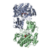 5nmwC  2xveS S: Starting model for refinement C: citing same article ( |
|---|---|
| Similar structure data |


-Links
 PDBj
PDBj- Assembly
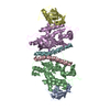
| Deposited unit | 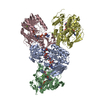
| ||||||||
|---|---|---|---|---|---|---|---|---|---|
| 1 | 
| ||||||||
| 2 | 
| ||||||||
| Unit cell |
|
-Components
| #1: Protein | Mass: 49299.051 Da / Num. of mol.: 4 Source method: isolated from a genetically manipulated source Source: (gene. exp.) Zonocerus variegatus (insect) / Gene: pno / Plasmid: pET28a+ / Production host:  #2: Chemical | ChemComp-FAD /   Mass: 785.550 Da / Num. of mol.: 4 / Source method: obtained synthetically / Formula: C27H33N9O15P2 / Comment: FAD*YM Mass: 785.550 Da / Num. of mol.: 4 / Source method: obtained synthetically / Formula: C27H33N9O15P2 / Comment: FAD*YM#3: Chemical | ChemComp-NAP /   Mass: 743.405 Da / Num. of mol.: 4 / Source method: obtained synthetically / Formula: C21H28N7O17P3 Mass: 743.405 Da / Num. of mol.: 4 / Source method: obtained synthetically / Formula: C21H28N7O17P3#4: Chemical |   Mass: 24.305 Da / Num. of mol.: 3 / Source method: obtained synthetically / Formula: Mg Mass: 24.305 Da / Num. of mol.: 3 / Source method: obtained synthetically / Formula: Mg#5: Water | ChemComp-HOH / |  Mass: 18.015 Da / Num. of mol.: 1572 / Source method: isolated from a natural source / Formula: H2O Mass: 18.015 Da / Num. of mol.: 1572 / Source method: isolated from a natural source / Formula: H2O |
|---|
-Experimental details
-Experiment
| Experiment | Method: X-RAY DIFFRACTION / Number of used crystals: 1 |
|---|
- Sample preparation
| Crystal | Density Matthews: 2.2 Å3/Da / Density % sol: 43.8 % |
|---|---|
| Crystal grow | Temperature: 291 K / Method: vapor diffusion, hanging drop / pH: 7 / Details: TRIS-HCl, MgCl2, PEG3350, NADP+ |
-Data collection
| Diffraction | Mean temperature: 100 K |
|---|---|
| Diffraction source | Source: SYNCHROTRON / Site: PETRA III, EMBL c/o DESY  / Beamline: P14 (MX2) / Wavelength: 0.9762 Å / Beamline: P14 (MX2) / Wavelength: 0.9762 Å |
| Detector | Type: DECTRIS PILATUS3 6M / Detector: PIXEL / Date: Oct 15, 2015 |
| Radiation | Protocol: SINGLE WAVELENGTH / Monochromatic (M) / Laue (L): M / Scattering type: x-ray |
| Radiation wavelength | Wavelength: 0.9762 Å / Relative weight: 1 |
| Reflection | Resolution: 1.6→77.13 Å / Num. obs: 213235 / % possible obs: 96.1 % / Redundancy: 4.9 % / Rmerge(I) obs: 0.081 / Net I/σ(I): 39.3 |
| Reflection shell | Resolution: 1.6→1.62 Å / Redundancy: 2.2 % / Rmerge(I) obs: 0.827 / Mean I/σ(I) all: 2.3 / % possible all: 47.5 |
- Processing
| Software |
| |||||||||||||||||||||
|---|---|---|---|---|---|---|---|---|---|---|---|---|---|---|---|---|---|---|---|---|---|---|
| Refinement | Method to determine structure: MOLECULAR REPLACEMENT Starting model: 2xve Resolution: 1.6→77.13 Å / Cor.coef. Fo:Fc: 0.965 / Cor.coef. Fo:Fc free: 0.953 / WRfactor Rfree: 0.1974 / WRfactor Rwork: 0.1704 / Occupancy max: 1 / Occupancy min: 0.13 / FOM work R set: 0.8716 / SU B: 0.002 / SU ML: 0 / SU R Cruickshank DPI: 0.0787 / SU Rfree: 0.0914 / Cross valid method: THROUGHOUT / σ(F): 0 / ESU R: 0.079 / ESU R Free: 0.091 / Stereochemistry target values: MAXIMUM LIKELIHOOD / Details: U VALUES : REFINED INDIVIDUALLY
| |||||||||||||||||||||
| Solvent computation | Ion probe radii: 0.8 Å / Shrinkage radii: 0.8 Å / VDW probe radii: 1.2 Å / Solvent model: MASK | |||||||||||||||||||||
| Displacement parameters | Biso max: 107.91 Å2 / Biso mean: 21.5286 Å2 / Biso min: 7 Å2
| |||||||||||||||||||||
| Refinement step | Cycle: LAST / Resolution: 1.6→77.13 Å
| |||||||||||||||||||||
| LS refinement shell | Resolution: 1.596→1.637 Å / Total num. of bins used: 20
|
