 Movie
Movie Controller

[English] 日本語
 Yorodumi
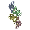
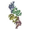
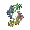
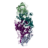
- PDB-5m4o: Crystal structure of hydroquinone 1,2-dioxygenase from Sphingomon... -
+ Open data

- Basic information
| Entry | Database: PDB / ID: 5m4o | ||||||
|---|---|---|---|---|---|---|---|
| Title | Crystal structure of hydroquinone 1,2-dioxygenase from Sphingomonas sp. TTNP3 in complex with 4-nitrophenol | ||||||
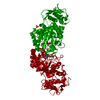
 Components Components |
| ||||||
 Keywords Keywords | OXIDOREDUCTASE / DIOXYGENASE / CUPIN | ||||||
| Function / homology |  Function and homology information Function and homology informationOxidoreductases; Acting on single donors with incorporation of molecular oxygen (oxygenases); With incorporation of two atoms of oxygen / dioxygenase activity / metal ion binding Similarity search - Function | ||||||
| Biological species |  Sphingomonas sp. TTNP3 (bacteria) Sphingomonas sp. TTNP3 (bacteria) | ||||||
| Method |  X-RAY DIFFRACTION / SYNCHROTRON / FOURIER SYNTHESIS / Resolution: 2.1 Å X-RAY DIFFRACTION / SYNCHROTRON / FOURIER SYNTHESIS / Resolution: 2.1 Å | ||||||
 Authors Authors | Ferraroni, M. / Da Vela, S. / Scozzafava, A. / Kolvenbach, B. / Corvini, P.F.X. | ||||||
 Citation Citation | Journal: Biochim. Biophys. Acta / Year: 2017 Title: The crystal structures of native hydroquinone 1,2-dioxygenase from Sphingomonas sp. TTNP3 and of substrate and inhibitor complexes. Authors: Ferraroni, M. / Da Vela, S. / Kolvenbach, B.A. / Corvini, P.F. / Scozzafava, A. | ||||||
| History |
|
- Structure visualization
| Structure viewer | Molecule: MolmilJmol/JSmol |
|---|
- Downloads & links
-Download
| PDBx/mmCIF format | 5m4o.cif.gz | 407.7 KB | Display | PDBx/mmCIF format |
|---|---|---|---|---|
| PDB format | pdb5m4o.ent.gz | 330.6 KB | Display | PDB format |
| PDBx/mmJSON format | 5m4o.json.gz | Tree view | PDBx/mmJSON format | |
| Others |  Other downloads Other downloads |
-Validation report
| Arichive directory | https://data.pdbj.org/pub/pdb/validation_reports/m4/5m4oftp://data.pdbj.org/pub/pdb/validation_reports/m4/5m4o | HTTPS FTP |
|---|
-Related structure data


-Links
 PDBj
PDBj
- Assembly
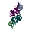
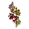
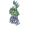
| Deposited unit | 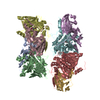
| ||||||||
|---|---|---|---|---|---|---|---|---|---|
| 1 | 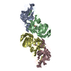
| ||||||||
| 2 | 
| ||||||||
| 3 | 
| ||||||||
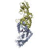
| Unit cell |
|
-Components
| #1: Protein | Mass: 18571.809 Da / Num. of mol.: 4 / Source method: isolated from a natural source / Details: Hydroquinone 1,2-dioxygenase small subunit / Source: (natural) Sphingomonas sp. TTNP3 (bacteria)References: UniProt: F8TW82, Oxidoreductases; Acting on single donors with incorporation of molecular oxygen (oxygenases); With incorporation of two atoms of oxygen #2: Protein | Mass: 38238.789 Da / Num. of mol.: 4 / Source method: isolated from a natural source / Details: Hydroquinone 1,2-dioxygenase large subunit / Source: (natural) Sphingomonas sp. TTNP3 (bacteria)References: UniProt: F8TW83, Oxidoreductases; Acting on single donors with incorporation of molecular oxygen (oxygenases); With incorporation of two atoms of oxygen #3: Chemical | ChemComp-FE /   Mass: 55.845 Da / Num. of mol.: 4 / Source method: obtained synthetically / Formula: Fe Mass: 55.845 Da / Num. of mol.: 4 / Source method: obtained synthetically / Formula: Fe#4: Chemical | ChemComp-NPO / 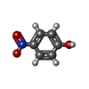  Mass: 139.109 Da / Num. of mol.: 4 / Source method: obtained synthetically / Formula: C6H5NO3 Mass: 139.109 Da / Num. of mol.: 4 / Source method: obtained synthetically / Formula: C6H5NO3#5: Water | ChemComp-HOH / |  Mass: 18.015 Da / Num. of mol.: 1076 / Source method: isolated from a natural source / Formula: H2O Mass: 18.015 Da / Num. of mol.: 1076 / Source method: isolated from a natural source / Formula: H2O |
|---|
-Experimental details
-Experiment
| Experiment | Method: X-RAY DIFFRACTION / Number of used crystals: 1 |
|---|
- Sample preparation
| Crystal | Density Matthews: 2.2 Å3/Da / Density % sol: 44.06 % |
|---|---|
| Crystal grow | Temperature: 296 K / Method: vapor diffusion / pH: 6.5 / Details: 14% PEG 3350, 0.35 M MgCl2 and 0.1 M Mes |
-Data collection
| Diffraction | Mean temperature: 100 K | ||||||||||||||||||||||||||||||||||||||||||||||||||||||||||||
|---|---|---|---|---|---|---|---|---|---|---|---|---|---|---|---|---|---|---|---|---|---|---|---|---|---|---|---|---|---|---|---|---|---|---|---|---|---|---|---|---|---|---|---|---|---|---|---|---|---|---|---|---|---|---|---|---|---|---|---|---|---|
| Diffraction source | Source: SYNCHROTRON / Site: ESRF  / Beamline: ID23-2 / Wavelength: 0.8729 Å / Beamline: ID23-2 / Wavelength: 0.8729 Å | ||||||||||||||||||||||||||||||||||||||||||||||||||||||||||||
| Detector | Type: MARMOSAIC 225 mm CCD / Detector: CCD / Date: May 12, 2016 | ||||||||||||||||||||||||||||||||||||||||||||||||||||||||||||
| Radiation | Protocol: SINGLE WAVELENGTH / Monochromatic (M) / Laue (L): M / Scattering type: x-ray | ||||||||||||||||||||||||||||||||||||||||||||||||||||||||||||
| Radiation wavelength | Wavelength: 0.8729 Å / Relative weight: 1 | ||||||||||||||||||||||||||||||||||||||||||||||||||||||||||||
| Reflection | Resolution: 2.1→29.7 Å / Num. obs: 112445 / % possible obs: 98.3 % / Observed criterion σ(I): -3 / Redundancy: 4.04 % / Biso Wilson estimate: 33.313 Å2 / CC1/2: 0.99 / Rmerge(I) obs: 0.161 / Net I/σ(I): 6.54 | ||||||||||||||||||||||||||||||||||||||||||||||||||||||||||||
| Reflection shell |
|
- Processing
| Software |
| ||||||||||||||||||||||||||||||||||||||||||||||||||||||||||||
|---|---|---|---|---|---|---|---|---|---|---|---|---|---|---|---|---|---|---|---|---|---|---|---|---|---|---|---|---|---|---|---|---|---|---|---|---|---|---|---|---|---|---|---|---|---|---|---|---|---|---|---|---|---|---|---|---|---|---|---|---|---|
| Refinement | Method to determine structure: FOURIER SYNTHESIS / Resolution: 2.1→29.697 Å / Cor.coef. Fo:Fc: 0.96 / Cor.coef. Fo:Fc free: 0.923 / SU B: 9.347 / SU ML: 0.22 / SU R Cruickshank DPI: 0.256 / Cross valid method: THROUGHOUT / σ(F): 0 / ESU R: 0.256 / ESU R Free: 0.21 / Details: U VALUES : REFINED INDIVIDUALLY
| ||||||||||||||||||||||||||||||||||||||||||||||||||||||||||||
| Solvent computation | Ion probe radii: 0.8 Å / Shrinkage radii: 0.8 Å / VDW probe radii: 1.2 Å | ||||||||||||||||||||||||||||||||||||||||||||||||||||||||||||
| Displacement parameters | Biso max: 99.25 Å2 / Biso mean: 33.931 Å2 / Biso min: 11.03 Å2
| ||||||||||||||||||||||||||||||||||||||||||||||||||||||||||||
| Refinement step | Cycle: final / Resolution: 2.1→29.697 Å
| ||||||||||||||||||||||||||||||||||||||||||||||||||||||||||||
| Refine LS restraints |
| ||||||||||||||||||||||||||||||||||||||||||||||||||||||||||||
| LS refinement shell | Resolution: 2.101→2.155 Å / Total num. of bins used: 20
|
