 Movie
Movie Controller


+ Open data

- Basic information
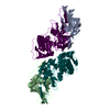
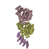
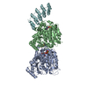
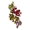
| Entry | Database: PDB / ID: 4zxd | ||||||
|---|---|---|---|---|---|---|---|
| Title | Crystal Structure of hydroquinone 1,2-dioxygenase PnpCD | ||||||
 Components Components |
| ||||||
 Keywords Keywords | OXIDOREDUCTASE / dioxygenase / cuipin / hydroquinone pathway | ||||||
| Function / homology | Hydroquinone 1,2-dioxygenase large subunit, N-terminal / Hydroquinone 1,2-dioxygenase large subunit N-terminal / dioxygenase activity / RmlC-like cupin domain superfamily / RmlC-like jelly roll fold / metal ion binding / Hydroquinone dioxygenase large subunit / Hydroquinone dioxygenase small subunit Function and homology information Function and homology information | ||||||
| Biological species |  Pseudomonas sp. (bacteria) Pseudomonas sp. (bacteria) | ||||||
| Method |  X-RAY DIFFRACTION / SYNCHROTRON / MOLECULAR REPLACEMENT / Resolution: 3.052 Å X-RAY DIFFRACTION / SYNCHROTRON / MOLECULAR REPLACEMENT / Resolution: 3.052 Å | ||||||
 Authors Authors | Liu, S. / Su, T. / Zhang, C. / Gu, L. | ||||||
| Funding support |  China, 1items China, 1items
| ||||||
 Citation Citation | Journal: J.Biol.Chem. / Year: 2015 Title: Crystal Structure of PnpCD, a Two-subunit Hydroquinone 1,2-Dioxygenase, Reveals a Novel Structural Class of Fe2+-dependent Dioxygenases. Authors: Liu, S. / Su, T. / Zhang, C. / Zhang, W.M. / Zhu, D. / Su, J. / Wei, T. / Wang, K. / Huang, Y. / Guo, L. / Xu, S. / Zhou, N.Y. / Gu, L. | ||||||
| History |
|
- Structure visualization
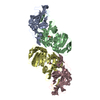
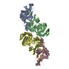
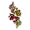
| Structure viewer | Molecule: MolmilJmol/JSmol |
|---|
- Downloads & links
-Download
| PDBx/mmCIF format | 4zxd.cif.gz | 199.6 KB | Display | PDBx/mmCIF format |
|---|---|---|---|---|
| PDB format | pdb4zxd.ent.gz | 158.7 KB | Display | PDB format |
| PDBx/mmJSON format | 4zxd.json.gz | Tree view | PDBx/mmJSON format | |
| Others |  Other downloads Other downloads |
-Validation report
| Arichive directory | https://data.pdbj.org/pub/pdb/validation_reports/zx/4zxdftp://data.pdbj.org/pub/pdb/validation_reports/zx/4zxd | HTTPS FTP |
|---|
-Related structure data
| Related structure data | 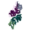 4zxaSC  4zxcC S: Starting model for refinement C: citing same article ( |
|---|---|
| Similar structure data |


-Links
 PDBj
PDBj- Assembly
| Deposited unit | 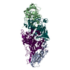
| ||||||||
|---|---|---|---|---|---|---|---|---|---|
| 1 |
| ||||||||
| Unit cell |
|
-Components
| #1: Protein | Mass: 18308.932 Da / Num. of mol.: 2 Source method: isolated from a genetically manipulated source Source: (gene. exp.) Pseudomonas sp. (strain WBC-3) (bacteria)Strain: WBC-3 / Plasmid: pET15b / Production host: #2: Protein | Mass: 38377.953 Da / Num. of mol.: 2 Source method: isolated from a genetically manipulated source Source: (gene. exp.) Pseudomonas sp. (strain WBC-3) (bacteria)Strain: WBC-3 / Plasmid: pET15b / Production host: #3: Water | ChemComp-HOH / |  Mass: 18.015 Da / Num. of mol.: 19 / Source method: isolated from a natural source / Formula: H2O Mass: 18.015 Da / Num. of mol.: 19 / Source method: isolated from a natural source / Formula: H2O |
|---|
-Experimental details
-Experiment
| Experiment | Method: X-RAY DIFFRACTION |
|---|
- Sample preparation
| Crystal | Density Matthews: 2.47 Å3/Da / Density % sol: 50.24 % |
|---|---|
| Crystal grow | Temperature: 293 K / Method: vapor diffusion, sitting drop / pH: 4.6 Details: 1.8 M Ammonium citrate dibasic, 0.1 M Sodium acetate pH 4.6 |
-Data collection
| Diffraction | Mean temperature: 100 K |
|---|---|
| Diffraction source | Source: SYNCHROTRON / Site: SSRF / Beamline: BL17U / Wavelength: 0.979 Å |
| Detector | Type: ADSC QUANTUM 315r / Detector: CCD / Date: Aug 12, 2013 |
| Radiation | Monochromator: Si (111) / Protocol: SINGLE WAVELENGTH / Monochromatic (M) / Laue (L): M / Scattering type: x-ray |
| Radiation wavelength | Wavelength: 0.979 Å / Relative weight: 1 |
| Reflection | Resolution: 3.05→50 Å / Num. obs: 21998 / % possible obs: 99.4 % / Redundancy: 6.6 % / Rsym value: 0.131 / Net I/σ(I): 19.3 |
| Reflection shell | Resolution: 3.05→3.16 Å / Redundancy: 6.4 % / Mean I/σ(I) obs: 4.6 / % possible all: 98.9 |
- Processing
| Software |
| |||||||||||||||||||||||||||||||||||||||||||||||||||||||||||||||
|---|---|---|---|---|---|---|---|---|---|---|---|---|---|---|---|---|---|---|---|---|---|---|---|---|---|---|---|---|---|---|---|---|---|---|---|---|---|---|---|---|---|---|---|---|---|---|---|---|---|---|---|---|---|---|---|---|---|---|---|---|---|---|---|---|
| Refinement | Method to determine structure: MOLECULAR REPLACEMENT Starting model: 4ZXA Resolution: 3.052→38.694 Å / SU ML: 0.33 / Cross valid method: FREE R-VALUE / σ(F): 1.34 / Phase error: 27.95 / Stereochemistry target values: ML
| |||||||||||||||||||||||||||||||||||||||||||||||||||||||||||||||
| Solvent computation | Shrinkage radii: 0.9 Å / VDW probe radii: 1.11 Å / Solvent model: FLAT BULK SOLVENT MODEL | |||||||||||||||||||||||||||||||||||||||||||||||||||||||||||||||
| Refinement step | Cycle: LAST / Resolution: 3.052→38.694 Å
| |||||||||||||||||||||||||||||||||||||||||||||||||||||||||||||||
| Refine LS restraints |
| |||||||||||||||||||||||||||||||||||||||||||||||||||||||||||||||
| LS refinement shell |
|
