 Movie
Movie Controller


+ Open data

- Basic information
| Entry | Database: PDB / ID: 6kbn | ||||||
|---|---|---|---|---|---|---|---|
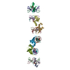
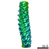
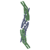
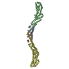
| Title | Crystal structure of Vac8 (del 19-33) bound to Atg13 | ||||||
 Components Components |
| ||||||
 Keywords Keywords | PROTEIN BINDING/PROTEIN TRANSPORT / Armadillo repeats / complex / PROTEIN BINDING-PROTEIN TRANSPORT complex | ||||||
| Function / homology |  Function and homology information Function and homology informationnucleus-vacuole junction assembly / Cvt vesicle assembly / PAS complex / Myo2p-Vac17p-Vac8p transport complex / lipophagy / protein localization to membrane raft / regulation of cellular localization / nucleus-vacuole junction / establishment of organelle localization / vacuole-isolation membrane contact site ...nucleus-vacuole junction assembly / Cvt vesicle assembly / PAS complex / Myo2p-Vac17p-Vac8p transport complex / lipophagy / protein localization to membrane raft / regulation of cellular localization / nucleus-vacuole junction / establishment of organelle localization / vacuole-isolation membrane contact site / armadillo repeat domain binding / protein targeting to vacuole involved in autophagy / vacuole inheritance / Macroautophagy / Atg1/ULK1 kinase complex / vacuole fusion, non-autophagic / cytoplasm to vacuole targeting by the Cvt pathway / nucleophagy / ribophagy / protein localization to phagophore assembly site / autophagy of mitochondrion / piecemeal microautophagy of the nucleus / pexophagy / protein kinase regulator activity / phagophore assembly site / fungal-type vacuole membrane / cellular response to nitrogen starvation / protein-containing complex localization / response to starvation / nuclear outer membrane / autophagosome assembly / mitophagy / protein-membrane adaptor activity / positive regulation of autophagy / autophagosome / lipid metabolic process / macroautophagy / autophagy / nuclear membrane / membrane raft / lipid binding / membrane / identical protein binding / cytosol Similarity search - Function | ||||||
| Biological species |  | ||||||
| Method |  X-RAY DIFFRACTION / SYNCHROTRON / MOLECULAR REPLACEMENT / Resolution: 3.2 Å X-RAY DIFFRACTION / SYNCHROTRON / MOLECULAR REPLACEMENT / Resolution: 3.2 Å | ||||||
 Authors Authors | Park, J. / Lee, C. | ||||||
 Citation Citation | Journal: Autophagy / Year: 2020 Title: Quaternary structures of Vac8 differentially regulate the Cvt and PMN pathways. Authors: Park, J. / Kim, H.I. / Jeong, H. / Lee, M. / Jang, S.H. / Yoon, S.Y. / Kim, H. / Park, Z.Y. / Jun, Y. / Lee, C. | ||||||
| History |
|
- Structure visualization
| Structure viewer | Molecule: MolmilJmol/JSmol |
|---|
- Downloads & links
-Download
| PDBx/mmCIF format | 6kbn.cif.gz | 419.8 KB | Display | PDBx/mmCIF format |
|---|---|---|---|---|
| PDB format | pdb6kbn.ent.gz | 348.3 KB | Display | PDB format |
| PDBx/mmJSON format | 6kbn.json.gz | Tree view | PDBx/mmJSON format | |
| Others |  Other downloads Other downloads |
-Validation report
| Arichive directory | https://data.pdbj.org/pub/pdb/validation_reports/kb/6kbnftp://data.pdbj.org/pub/pdb/validation_reports/kb/6kbn | HTTPS FTP |
|---|
-Related structure data
| Related structure data |  6kbmC  5xjgS S: Starting model for refinement C: citing same article ( |
|---|---|
| Similar structure data |

-Links
 PDBj
PDBj


- Assembly
| Deposited unit | 
| ||||||||
|---|---|---|---|---|---|---|---|---|---|
| 1 |
| ||||||||
| Unit cell |
|
-Components
| #1: Protein | Mass: 61676.777 Da / Num. of mol.: 2 / Mutation: residues 19-33 deletion Source method: isolated from a genetically manipulated source Source: (gene. exp.) Production host:  #2: Protein | Mass: 14249.582 Da / Num. of mol.: 2 Source method: isolated from a genetically manipulated source Source: (gene. exp.) Production host: |
|---|
-Experimental details
-Experiment
| Experiment | Method: X-RAY DIFFRACTION / Number of used crystals: 1 |
|---|
- Sample preparation
| Crystal | Density Matthews: 2.66 Å3/Da / Density % sol: 53.77 % |
|---|---|
| Crystal grow | Temperature: 277 K / Method: vapor diffusion, hanging drop Details: PEG 3350, N-(2-Acetamido)iminodiacetic acid, Ammonium sulfate |
-Data collection
| Diffraction | Mean temperature: 100 K / Serial crystal experiment: N |
|---|---|
| Diffraction source | Source: SYNCHROTRON / Site: PAL/PLS  / Beamline: 5C (4A) / Wavelength: 0.97949 Å / Beamline: 5C (4A) / Wavelength: 0.97949 Å |
| Detector | Type: DECTRIS PILATUS3 6M / Detector: PIXEL / Date: May 9, 2017 |
| Radiation | Protocol: SINGLE WAVELENGTH / Monochromatic (M) / Laue (L): M / Scattering type: x-ray |
| Radiation wavelength | Wavelength: 0.97949 Å / Relative weight: 1 |
| Reflection | Resolution: 3.2→50 Å / Num. obs: 27903 / % possible obs: 98.7 % / Redundancy: 3.8 % / Rmerge(I) obs: 0.103 / Net I/σ(I): 17.88 |
| Reflection shell | Resolution: 3.2→3.26 Å / Rmerge(I) obs: 0.537 / Num. unique obs: 1320 |
- Processing
| Software |
| |||||||||||||||||||||||||||||||||||||||||||||||||||||||||||||||||||||||||||||||||||||||||||||||||||||||||||||||||||||||||||||
|---|---|---|---|---|---|---|---|---|---|---|---|---|---|---|---|---|---|---|---|---|---|---|---|---|---|---|---|---|---|---|---|---|---|---|---|---|---|---|---|---|---|---|---|---|---|---|---|---|---|---|---|---|---|---|---|---|---|---|---|---|---|---|---|---|---|---|---|---|---|---|---|---|---|---|---|---|---|---|---|---|---|---|---|---|---|---|---|---|---|---|---|---|---|---|---|---|---|---|---|---|---|---|---|---|---|---|---|---|---|---|---|---|---|---|---|---|---|---|---|---|---|---|---|---|---|---|
| Refinement | Method to determine structure: MOLECULAR REPLACEMENT Starting model: 5XJG Resolution: 3.2→35.113 Å / SU ML: 0.41 / Cross valid method: FREE R-VALUE / σ(F): 1.4 / Phase error: 24.96
| |||||||||||||||||||||||||||||||||||||||||||||||||||||||||||||||||||||||||||||||||||||||||||||||||||||||||||||||||||||||||||||
| Solvent computation | Shrinkage radii: 0.9 Å / VDW probe radii: 1.11 Å | |||||||||||||||||||||||||||||||||||||||||||||||||||||||||||||||||||||||||||||||||||||||||||||||||||||||||||||||||||||||||||||
| Refinement step | Cycle: LAST / Resolution: 3.2→35.113 Å
| |||||||||||||||||||||||||||||||||||||||||||||||||||||||||||||||||||||||||||||||||||||||||||||||||||||||||||||||||||||||||||||
| Refine LS restraints |
| |||||||||||||||||||||||||||||||||||||||||||||||||||||||||||||||||||||||||||||||||||||||||||||||||||||||||||||||||||||||||||||
| LS refinement shell |
| |||||||||||||||||||||||||||||||||||||||||||||||||||||||||||||||||||||||||||||||||||||||||||||||||||||||||||||||||||||||||||||
| Refinement TLS params. | Method: refined / Refine-ID: X-RAY DIFFRACTION
| |||||||||||||||||||||||||||||||||||||||||||||||||||||||||||||||||||||||||||||||||||||||||||||||||||||||||||||||||||||||||||||
| Refinement TLS group |
|
